Cat.No:GM-49245AB
Product:Anti-H_TNFRSF1B(TNFR2) hIgG1 Antibody(UC2.3.8)
Cat.No:GM-49245AB
Product:Anti-H_TNFRSF1B(TNFR2) hIgG1 Antibody(UC2.3.8)
GM-49245AB-10 10 μg
GM-49245AB-100 100 μg
GM-49245AB-1000 1 mg
Species Reactivity Human
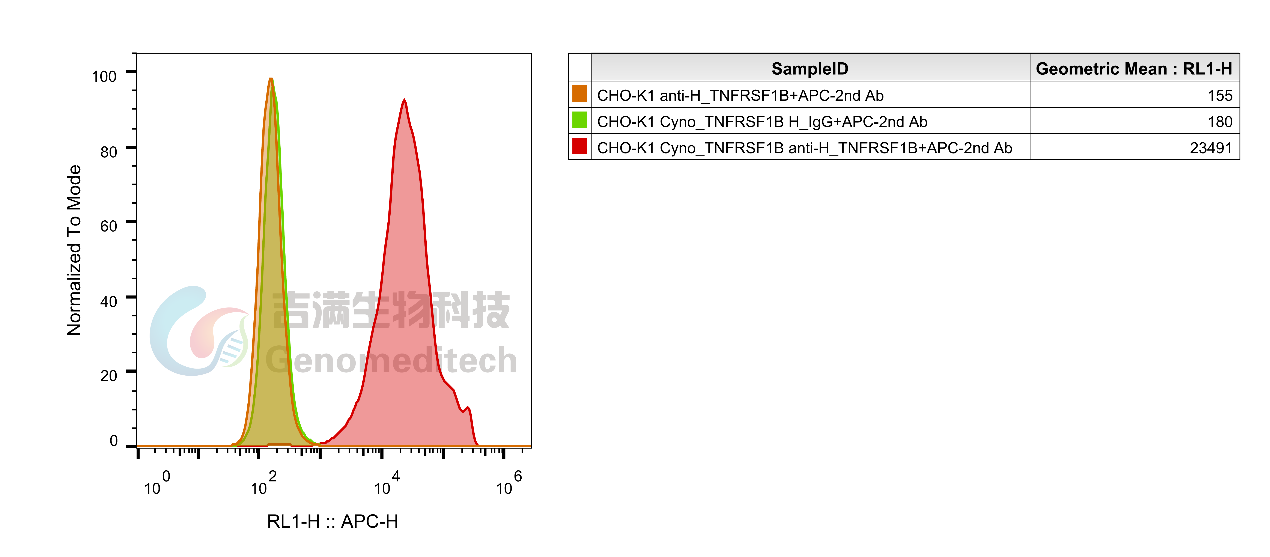
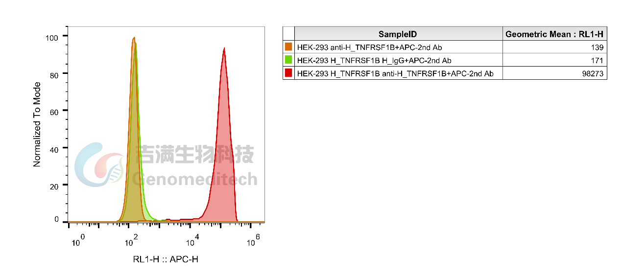
Specificity Detects human TNFRSF1B. It also detects cynomolgus TNFRSF1B.
Source/Isotype Monoclonal human IgG1, κ
Other Names p75; TBPII; TNFBR; CD120b; TNFR1B; TNFR80; TNF-R75; p75TNFR; TNF-R-II
Gene ID 7133(human), 102144224(cynomolgus)
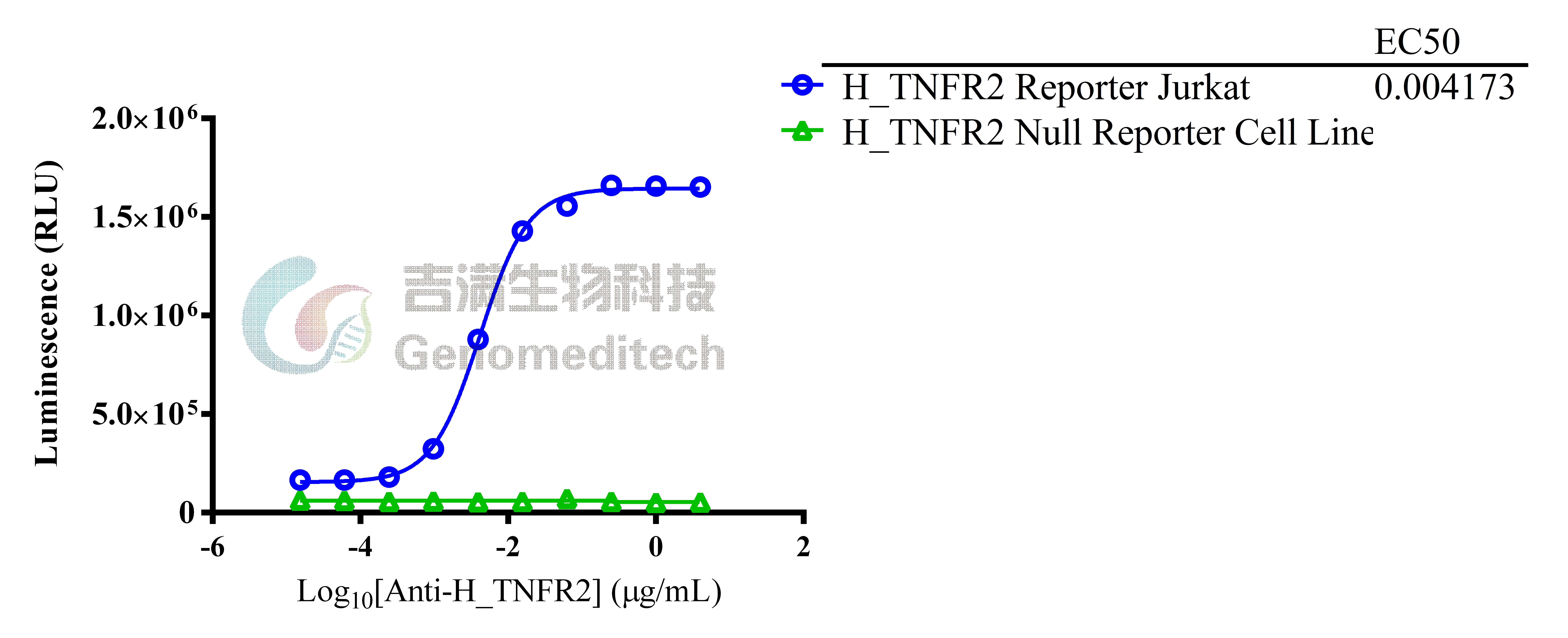
Application Flow cytometry: 1 μg-4 μg /1E6 cells;FC-Quality tested;Binding activation: 14 pg/mL-15 μg/mL
Background Tumor necrosis factor receptor superfamily member 1B (TNFRSF1B), also known as tumor necrosis factor receptor 2 (TNFR2) and CD120b, is one of two membrane receptors that binds tumor necrosis factor-alpha (TNFα). Like its counterpart, tumor necrosis factor receptor 1 (TNFR1), the extracellular region of TNFR2 consists of four cysteine-rich domains which allow for binding to TNFα. TNFR1 and TNFR2 possess different functions when bound to TNFα due to differences in their intracellular structures, such as TNFR2 lacking a death domain (DD). The protein encoded by this gene is a member of the tumor necrosis factor receptor superfamily, which also contains TNFRSF1A. This protein and TNF-receptor 1 form a heterocomplex that mediates the recruitment of two anti-apoptotic proteins, c-IAP1 and c-IAP2, which possess E3 ubiquitin ligase activity. The function of IAPs in TNF-receptor signalling is unknown, however, c-IAP1 is thought to potentiate TNF-induced apoptosis by the ubiquitination and degradation of TNF-receptor-associated factor 2 (TRAF2), which mediates anti-apoptotic signals. Knockout studies in mice also suggest a role of this protein in protecting neurons from apoptosis by stimulating antioxidative pathways.
Storage Store at +4℃ short term (1-2 weeks). Store at -20℃ long term
Formulation Phosphate-buffered solution, pH 7.2.


![]()

Cat.No:GM-49245AB
Product:Anti-H_TNFRSF1B(TNFR2) hIgG1 Antibody(UC2.3.8)
GM-49245AB-10 10 μg
GM-49245AB-100 100 μg
GM-49245AB-1000 1 mg
Species Reactivity Human
Specificity Detects human TNFRSF1B. It also detects cynomolgus TNFRSF1B.
Source/Isotype Monoclonal human IgG1, κ
Other Names p75; TBPII; TNFBR; CD120b; TNFR1B; TNFR80; TNF-R75; p75TNFR; TNF-R-II
Gene ID 7133(human), 102144224(cynomolgus)
Application Flow cytometry: 1 μg-4 μg /1E6 cells;FC-Quality tested;Binding activation: 14 pg/mL-15 μg/mL
Background Tumor necrosis factor receptor superfamily member 1B (TNFRSF1B), also known as tumor necrosis factor receptor 2 (TNFR2) and CD120b, is one of two membrane receptors that binds tumor necrosis factor-alpha (TNFα). Like its counterpart, tumor necrosis factor receptor 1 (TNFR1), the extracellular region of TNFR2 consists of four cysteine-rich domains which allow for binding to TNFα. TNFR1 and TNFR2 possess different functions when bound to TNFα due to differences in their intracellular structures, such as TNFR2 lacking a death domain (DD). The protein encoded by this gene is a member of the tumor necrosis factor receptor superfamily, which also contains TNFRSF1A. This protein and TNF-receptor 1 form a heterocomplex that mediates the recruitment of two anti-apoptotic proteins, c-IAP1 and c-IAP2, which possess E3 ubiquitin ligase activity. The function of IAPs in TNF-receptor signalling is unknown, however, c-IAP1 is thought to potentiate TNF-induced apoptosis by the ubiquitination and degradation of TNF-receptor-associated factor 2 (TRAF2), which mediates anti-apoptotic signals. Knockout studies in mice also suggest a role of this protein in protecting neurons from apoptosis by stimulating antioxidative pathways.
Storage Store at +4℃ short term (1-2 weeks). Store at -20℃ long term
Formulation Phosphate-buffered solution, pH 7.2.


![]()
