Cat,No:GM-28206AB
Product :Anti-mouse_PD1 mIgG1 Antibody(RMP1-14)
Cat,No:GM-28206AB
Product :Anti-mouse_PD1 mIgG1 Antibody(RMP1-14)
GM-28206AB-1mg / 1 mg
GM-28206AB-5mg / 5 mg
GM-28206AB-25mg / 5 mg*5 vials
GM-28206AB-50mg / 50 mg
GM-28206AB-100mg / 50 mg*2 vials
Expression System CHO
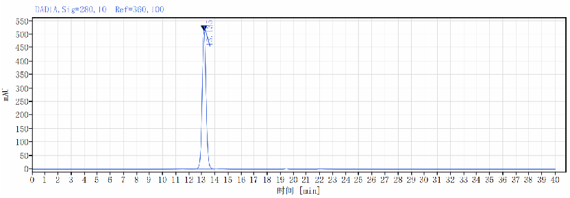
Aggregation < 5% as determined by SEC-HPLC
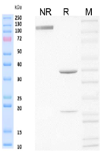
Purity >95% as determined by SDS-PAGE
Endotoxin <1 EU/mg, determined by LAL gel clotting assay
Sterility 0.2 μm Filtered
Target PD1
Clone RMP1-14
Alternative Names PD-1; Pdc1; Ly101
Sourcellsotype Monoclonal Mouse IgG1, κ
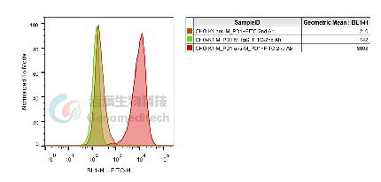
Application Flow cytometry; Binding activation: 1.5 ng/mL-100 μg/mL
Description The programmed cell death 1 protein (PD-1, PDCD1, CD279) is a member of the CD28 family of immunoreceptors that regulate T cell activation and immune responses. The PD-1 protein contains an extracellular Ig V domain, a transmembrane domain, and a cytoplasmic tail that includes an immunoreceptor tyrosine-based inhibitory motif (ITIM) and an immunoreceptor tyrosine-based switch motif (ITSM). PD-1 is activated by the cell surface ligands PD-L1 and PD-L2. Upon activation, PD-1 ITIM and ITSM phosphorylation leads to the recruitment of the protein tyrosine phosphatases SHP-1 and SHP-2, which suppress TCR signaling. In addition to activated T-cells, PD-1 is expressed in activated B-cells and monocytes, although its function in these cell types has not been fully characterized. The PD-1 pathway plays an important role in immune tolerance; however, research studies show that cancer cells often adopt this pathway to escape immune surveillance. Consequently, blockade of PD-1 and its ligands is proving to be a sound strategy for neoplastic intervention.
Formulation Phosphate-buffered solution, pH 7.2-7.4.


