Cat. No:GM-032AS012
Product:H_LAG3 Reporter Blockade Assay (Raji)

Cat. No:GM-032AS012
Product:H_LAG3 Reporter Blockade Assay (Raji)

H_LAG3 Reporter Jurkat Cell Line
Cell Growth Medium:RPMI 1640+10% FBS+1% P.S+3.5 μg/mL Blasticidin+400 μg/mL G418+0.75 μg/mL Puromycin
Cell Freezing Medium:90% FBS+10% DMSO
Raji Cell Line
Cell Growth Medium:RPMI 1640+10% FBS+1% P.S
Cell Freezing Medium:90% FBS+10% DMSO
Assay Buffer:RPMI 1640+1% FBS+1% P.S
Lymphocyte activation gene-3 (LAG-3; CD223) is a type I transmembrane protein expressed on the surface of activated CD4+ and CD8+ T cells, as well as NK and dendritic cell subsets. LAG-3 is closely related to CD4 and both molecules have four extracellular Ig-like domains, but LAG-3 has a much higher affinity for MHC II compared to CD4, approximately 100 times higher. The conventional view is that blocking the interaction between LAG-3 and MHC II can restore T cell activity.
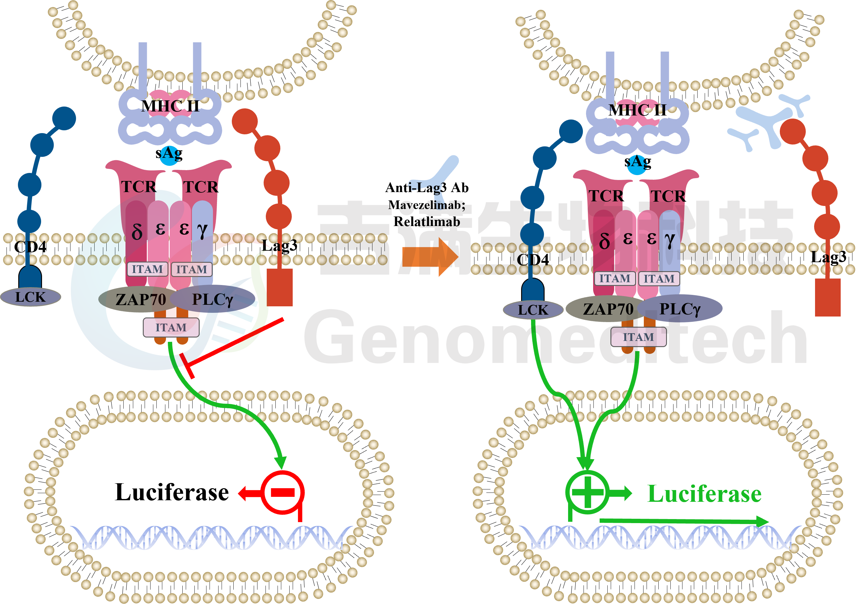
The JIMIN Bioscience H_LAG3 Reporter Blockade Assay reporter gene cell line is a Luciferase reporter gene cell line constructed based on the Lag3 signaling pathway. These cells stably express Lag3 gene, Luciferase reporter gene, and endogenously express CD4. By co-culturing these cells with the Raji Cell Line (which endogenously expresses MHC II molecules) and then adding sAg to stimulate T cell signals, the presence of Lag3 blocks the binding of MHC II molecules with CD4, thereby inhibiting T cell signal. By adding Anti-Lag3 Antibody, the interaction between Lag3 and MHC II molecules is blocked, restoring T cell signal. Therefore, this assay can be used to study Lag3-related antagonists as drugs.
Cat. No:GM-032AS012
Product:H_LAG3 Reporter Blockade Assay (Raji)

H_LAG3 Reporter Jurkat Cell Line
Cell Growth Medium:RPMI 1640+10% FBS+1% P.S+3.5 μg/mL Blasticidin+400 μg/mL G418+0.75 μg/mL Puromycin
Cell Freezing Medium:90% FBS+10% DMSO
Raji Cell Line
Cell Growth Medium:RPMI 1640+10% FBS+1% P.S
Cell Freezing Medium:90% FBS+10% DMSO
Assay Buffer:RPMI 1640+1% FBS+1% P.S
Lymphocyte activation gene-3 (LAG-3; CD223) is a type I transmembrane protein expressed on the surface of activated CD4+ and CD8+ T cells, as well as NK and dendritic cell subsets. LAG-3 is closely related to CD4 and both molecules have four extracellular Ig-like domains, but LAG-3 has a much higher affinity for MHC II compared to CD4, approximately 100 times higher. The conventional view is that blocking the interaction between LAG-3 and MHC II can restore T cell activity.
The JIMIN Bioscience H_LAG3 Reporter Blockade Assay reporter gene cell line is a Luciferase reporter gene cell line constructed based on the Lag3 signaling pathway. These cells stably express Lag3 gene, Luciferase reporter gene, and endogenously express CD4. By co-culturing these cells with the Raji Cell Line (which endogenously expresses MHC II molecules) and then adding sAg to stimulate T cell signals, the presence of Lag3 blocks the binding of MHC II molecules with CD4, thereby inhibiting T cell signal. By adding Anti-Lag3 Antibody, the interaction between Lag3 and MHC II molecules is blocked, restoring T cell signal. Therefore, this assay can be used to study Lag3-related antagonists as drugs.