Cat. No:GM-31740AB
Product:Anti-H_CD274(PDL1) hIgG1 Antibody(Atezolizumab)
Cat. No:GM-31740AB
Product:Anti-H_CD274(PDL1) hIgG1 Antibody(Atezolizumab)
GM-31740AB-10 10 μg
GM-31740AB-100 100 μg
GM-31740AB-1000 1mg
Species Reactivity Human; Mouse
Clone Atezolizumab
Source/Isotype Monoclonal Human IgG1 /κ
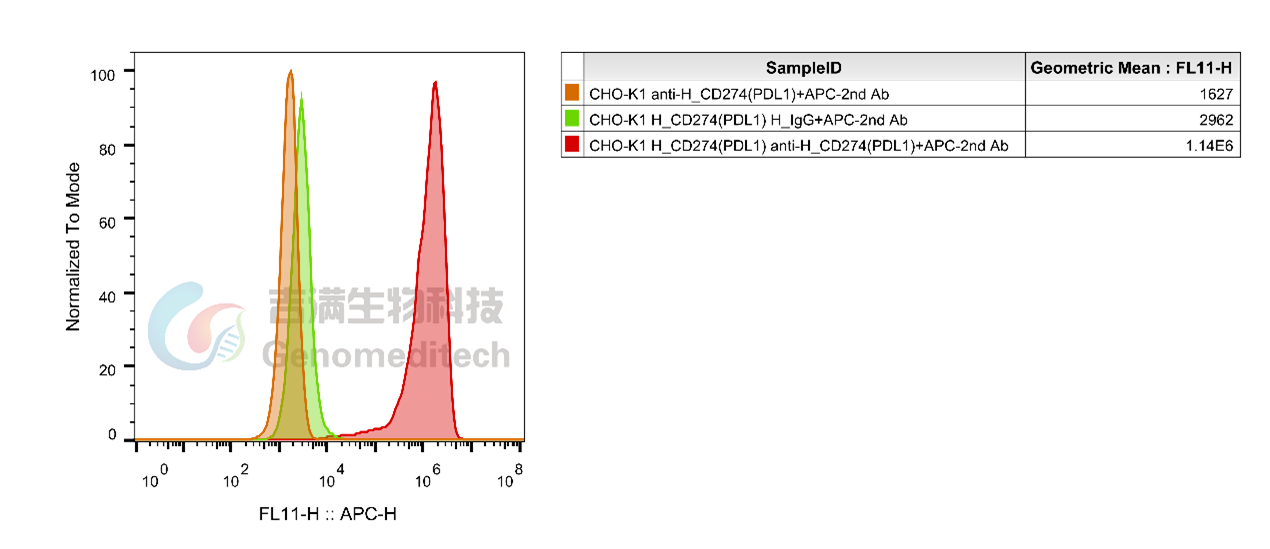
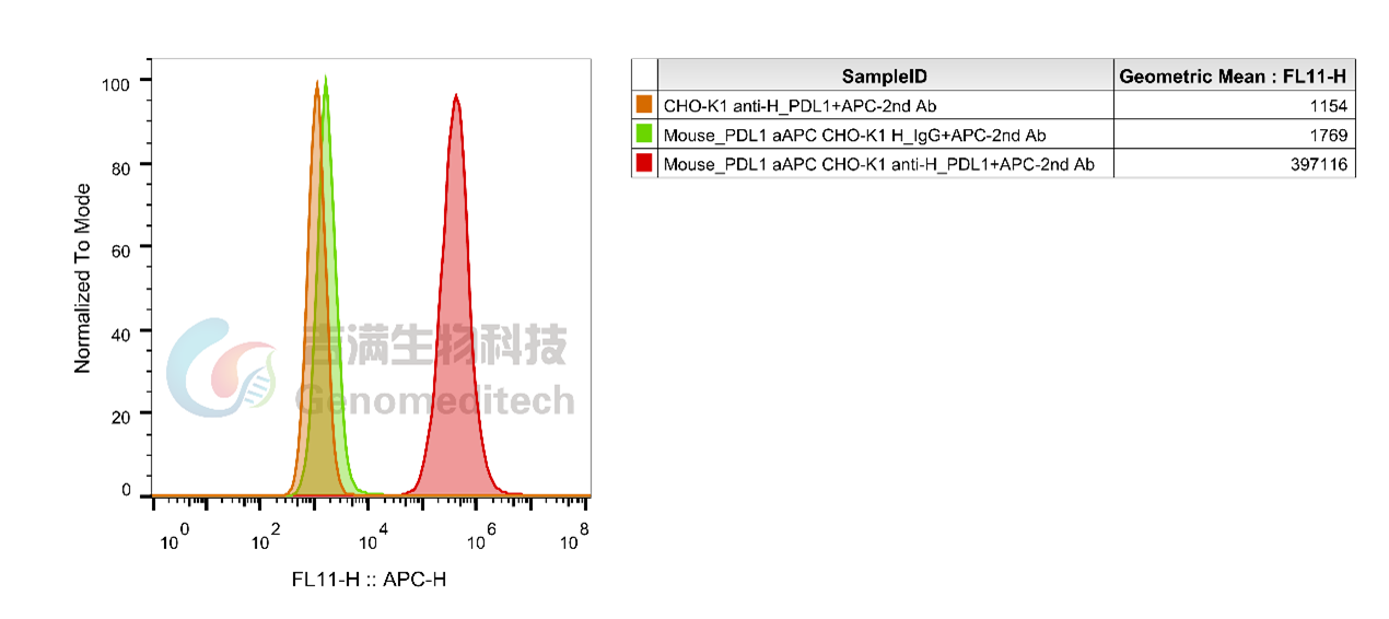
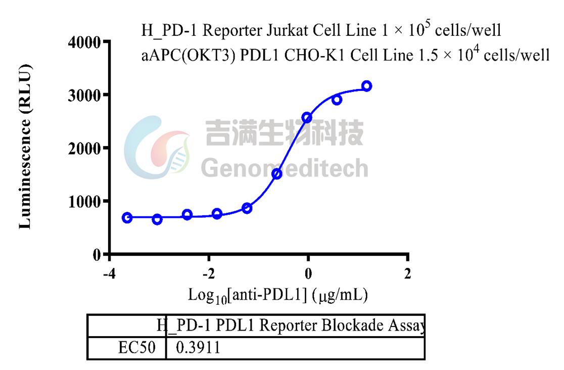
Application Flow Cytometry; Block assay
Specificity Detects PDL1
Gene PDL1
Other Names B7-H, B7H1, PD-L1, PDCD1L1, PDCD1LG1, CD274, hPD-L1
Gene ID 29126(human), 60533(mouse)
Background Programmed death-ligand 1 (PD-L1) also known as cluster of differentiation 274 (CD274) or B7 homolog 1 (B7-H1) is a protein that in humans is encoded by the CD274 gene. Programmed death-ligand 1 (PD-L1) is a 40kDa type 1 transmembrane protein that has been speculated to play a major role in suppressing the adaptive arm of immune systems during particular events such as pregnancy, tissue allografts, autoimmune disease and other disease states such as hepatitis. Normally the adaptive immune system reacts to antigens that are associated with immune system activation by exogenous or endogenous danger signals. In turn, clonal expansion of antigen-specific CD8+ T cells and/or CD4+ helper cells is propagated. The binding of PD-L1 to the inhibitory checkpoint molecule PD-1 transmits an inhibitory signal based on interaction with phosphatases (SHP-1 or SHP-2) via Immunoreceptor Tyrosine-Based Switch Motif (ITSM). This reduces the proliferation of antigen-specific T-cells in lymph nodes, while simultaneously reducing apoptosis in regulatory T cells (anti-inflammatory, suppressive T cells) - further mediated by a lower regulation of the gene Bcl-2.
Storage Store at 2-8℃ short term (1-2 weeks).Store at ≤ -20℃ long term. Avoid repeated freeze-thaw.
Formulation Phosphate-buffered solution, pH 7.2.
Endotoxin < 1 EU/mg, determined by LAL gel clotting assay


