Tumor Necrosis Factor (TNF) is a pro-inflammatory cytokine that coordinates tissue homeostasis by synergistically regulating the production of other cytokines, cell survival & death. In 1975, Carswell et al. discovered that after injecting lipopolysaccharide (LPS) into mice previously inoculated with Bacillus Calmette-Guérin (BCG), a substance capable of inducing tumor hemorrhagic necrosis appeared in the serum, which was subsequently named tumor necrosis factor (TNF). Moving into the 1980s, research confirmed that TNF plays a crucial role in cachexia and established that it is primarily produced by activated macrophages, NK cells, and T lymphocytes. In 1985, Shalaby named the macrophage-derived TNF as TNF-α and the T-lymphocyte-derived lymphotoxin as TNF-β to distinguish their origins and functional differences.
The Classification of TNF
TNF is classified into two types: TNF-α and TNF-β. Although TNF-α and TNF-β share only about 30% homology, they bind to the same receptors. Since the biological activity of TNF-α accounts for 70% to 95% of the total TNF activity, the term TNF as commonly used today typically refers to TNF-α.
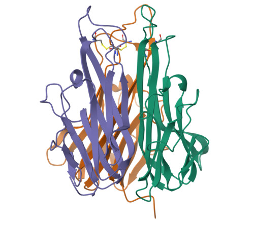
TNF-α (Tumor Necrosis Factor α) is a homotrimeric protein composed of 157 amino acids. It is primarily released and produced by activated macrophages and other cell types, such as CD4+ T cells, neutrophils, and mast cells. Capable of inducing inflammatory responses and apoptosis, it plays a vital role in multiple physiological processes, including immune responses, inflammation, and programmed cell death.
TNF-β (Tumor Necrosis Factor β), also known as lymphotoxin-α, is a cytokine produced by lymphocytes. TNF-β consists of 205 amino acid residues, including a 34-amino-acid signal peptide. The mature TNF-β molecule contains 171 amino acid residues and has a molecular weight of approximately 25 kDa.
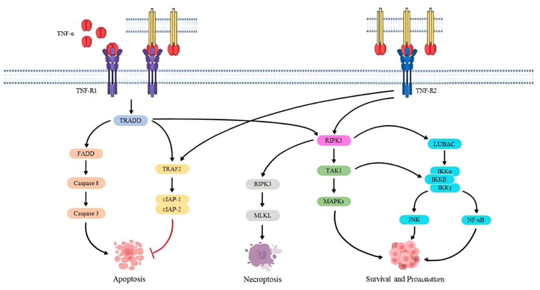
Receptor Signaling of TNF-α
TNF Pathway
The biological functions of TNF depend on the activation of downstream signaling pathways upon binding to its receptors. The primary receptors for TNF include TNFR1 (TNF Receptor 1) and TNFR2 (TNF Receptor 2), which exhibit significant differences in structure, expression distribution, and regulatory functions.
Structural Diagram of TNF-α
We Provide:
To meet the demands for drugs targeting the TNF/TNFR pathway, Genomeditech provides stable overexpressing cell lines, reporter gene cell lines, antibodies, and protein products to comprehensively accelerate your project progression.
1. Broad Species Coverage: Off-the-shelf products include both human- and monkey-derived TNFR2 overexpressing cell lines.
2. High Pathway Specificity: Equipped with a variety of reporter gene cell lines, our products meet diverse experimental design requirements and flexibly adapt to different R&D scenarios.
3. Rich Application Scenarios: Covering applications such as antibody functional evaluation, providing a more authentic approach for in vitro efficacy assessment.
4. Comprehensive Experimental Quality Control: Accompanied by matching Null control cell lines, effectively eliminating background signal interference to ensure the accuracy and reliability of experimental results.





| Product No. | Product Name | Gene | Pre-order/order |
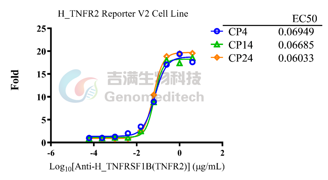
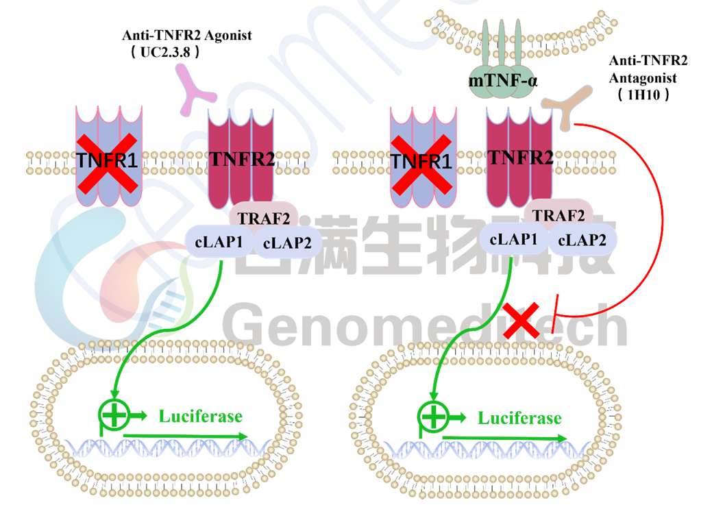
GM H_TNFR2 Reporter V2 Cell Line is a luciferase reporter cell line with TNFR1 knocked out.
|
Functional Validation
|
Stability Validation
|
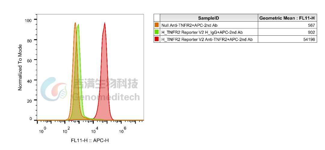
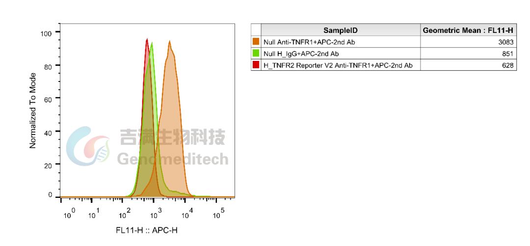
Flow Cytometry Validation: Flow cytometry verification confirms that the H_TNFR2 Reporter V2 Cell Line expresses only TNFR2, with no expression of TNFR1. This line is highly suitable for studying the signaling pathways between TNF-α and TNFR2, as well as for related drug discovery and development.
 |
 |

Tumor Necrosis Factor (TNF) is a pro-inflammatory cytokine that coordinates tissue homeostasis by synergistically regulating the production of other cytokines, cell survival & death. In 1975, Carswell et al. discovered that after injecting lipopolysaccharide (LPS) into mice previously inoculated with Bacillus Calmette-Guérin (BCG), a substance capable of inducing tumor hemorrhagic necrosis appeared in the serum, which was subsequently named tumor necrosis factor (TNF). Moving into the 1980s, research confirmed that TNF plays a crucial role in cachexia and established that it is primarily produced by activated macrophages, NK cells, and T lymphocytes. In 1985, Shalaby named the macrophage-derived TNF as TNF-α and the T-lymphocyte-derived lymphotoxin as TNF-β to distinguish their origins and functional differences.
The Classification of TNF
TNF is classified into two types: TNF-α and TNF-β. Although TNF-α and TNF-β share only about 30% homology, they bind to the same receptors. Since the biological activity of TNF-α accounts for 70% to 95% of the total TNF activity, the term TNF as commonly used today typically refers to TNF-α.
TNF-α (Tumor Necrosis Factor α) is a homotrimeric protein composed of 157 amino acids. It is primarily released and produced by activated macrophages and other cell types, such as CD4+ T cells, neutrophils, and mast cells. Capable of inducing inflammatory responses and apoptosis, it plays a vital role in multiple physiological processes, including immune responses, inflammation, and programmed cell death.
TNF-β (Tumor Necrosis Factor β), also known as lymphotoxin-α, is a cytokine produced by lymphocytes. TNF-β consists of 205 amino acid residues, including a 34-amino-acid signal peptide. The mature TNF-β molecule contains 171 amino acid residues and has a molecular weight of approximately 25 kDa.
Receptor Signaling of TNF-α
TNF Pathway
The biological functions of TNF depend on the activation of downstream signaling pathways upon binding to its receptors. The primary receptors for TNF include TNFR1 (TNF Receptor 1) and TNFR2 (TNF Receptor 2), which exhibit significant differences in structure, expression distribution, and regulatory functions.
Structural Diagram of TNF-α
We Provide:
To meet the demands for drugs targeting the TNF/TNFR pathway, Genomeditech provides stable overexpressing cell lines, reporter gene cell lines, antibodies, and protein products to comprehensively accelerate your project progression.
1. Broad Species Coverage: Off-the-shelf products include both human- and monkey-derived TNFR2 overexpressing cell lines.
2. High Pathway Specificity: Equipped with a variety of reporter gene cell lines, our products meet diverse experimental design requirements and flexibly adapt to different R&D scenarios.
3. Rich Application Scenarios: Covering applications such as antibody functional evaluation, providing a more authentic approach for in vitro efficacy assessment.
4. Comprehensive Experimental Quality Control: Accompanied by matching Null control cell lines, effectively eliminating background signal interference to ensure the accuracy and reliability of experimental results.
| Product No. | Product Name | Gene | Pre-order/order |
GM H_TNFR2 Reporter V2 Cell Line is a luciferase reporter cell line with TNFR1 knocked out.
|
Functional Validation
|
Stability Validation
|
Flow Cytometry Validation: Flow cytometry verification confirms that the H_TNFR2 Reporter V2 Cell Line expresses only TNFR2, with no expression of TNFR1. This line is highly suitable for studying the signaling pathways between TNF-α and TNFR2, as well as for related drug discovery and development.
|
|