
Tumor immunotherapy has become an indispensable pillar of modern cancer treatment. The success of these therapies largely depends on the recognition of tumor antigens by T cells and the subsequent activation of the immune response.
Tumor antigens are generally divided into two categories: Tumor-Specific Antigens (TSAs) and Tumor-Associated Antigens (TAAs). TSAs are exclusively expressed on tumor cells and are typically absent or expressed at extremely low levels in normal cells, limiting their widespread clinical application. Conversely, TAAs usually arise from gene amplification, aberrant genetic regulation, or post-translational modifications. They exhibit high sharing and prevalence across the same tumor types among different patients.
TAAs are typically overexpressed on the surface of tumor cells while showing trace or selective expression in normal host tissues. When targeting specific malignancies, TAAs serve as highly promising therapeutic targets. They are now widely applied in the treatment of various tumors and play a critical role in the research and development of human oncology drugs. Antibodies targeting TAAs can not only mediate tumor cell killing through effector mechanisms such as ADCC (Antibody-Dependent Cellular Cytotoxicity) and CDC (Complement-Dependent Cytotoxicity), but can also serve as diagnostic biomarkers or be utilized to enhance the tumor-targeting specificity of traditional therapies.
Characteristics of TAAs
- Expressed not only in tumor cells but also partially present in normal host cells.
- Primarily driven by gene amplification, aberrant genetic regulation, or post-translational modifications, leading to upregulated expression in tumor cells.
- Exhibiting significantly higher expression levels in tumor cells compared to normal cells.
- Serving as diagnostic biomarkers or immunotherapy targets; they are critical targets for oncology immunotherapy strategies such as monoclonal antibodies, ADCs (Antibody-Drug Conjugates), and CAR-T (Chimeric Antigen Receptor T-cell) therapies.
We provide:
To support the research and development of tumor immunotherapy drugs, Genomeditech stays at the forefront of global biomedical R&D, meticulously building a comprehensive product library of TAA-related tool cell lines.
We currently offer 350+ off-the-shelf TAA-related products, covering 80+ target genes. These products faithfully recapitulate the presentation and native conformation of antigens on the cell surface, accelerating your drug screening and efficacy evaluation workflows.
|
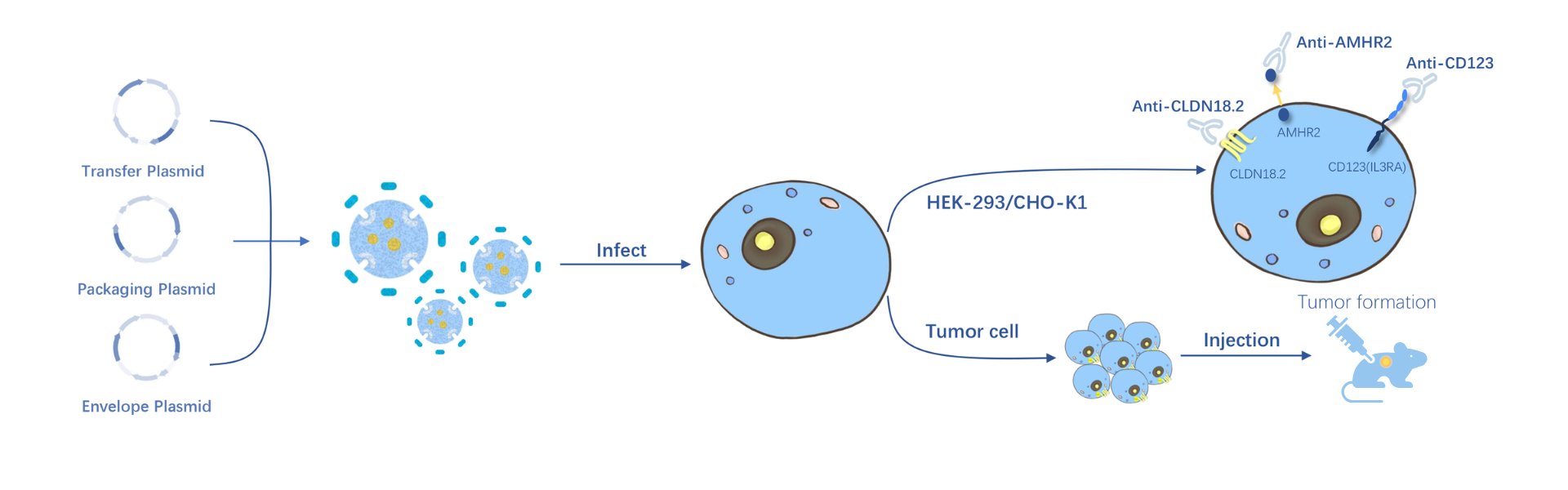
Stable Cell Lines Based on HEK293 or CHO-K1 Backgrounds These cell lines, which stably express specific antigens, are primarily used for antibody-specific binding assays, affinity testing, and in vitro functional screening against relevant targets (e.g., CLDN18.2, AMHR2, CD123). |
Luciferase-Labeled Stable Cell Lines Based on Tumor Backgrounds These lines can be inoculated into mice to form solid tumors for the construction of CDX mouse models. Combined with in vivo optical imaging technology, they enable real-time, dynamic monitoring of tumor growth and metastasis in vivo. Widely utilized in in vivo efficacy evaluation, CAR-T cell therapy assessment, and the validation of other anti-tumor therapeutics. |
| Product No. | Product Name | Gene | Pre-order/order |
|
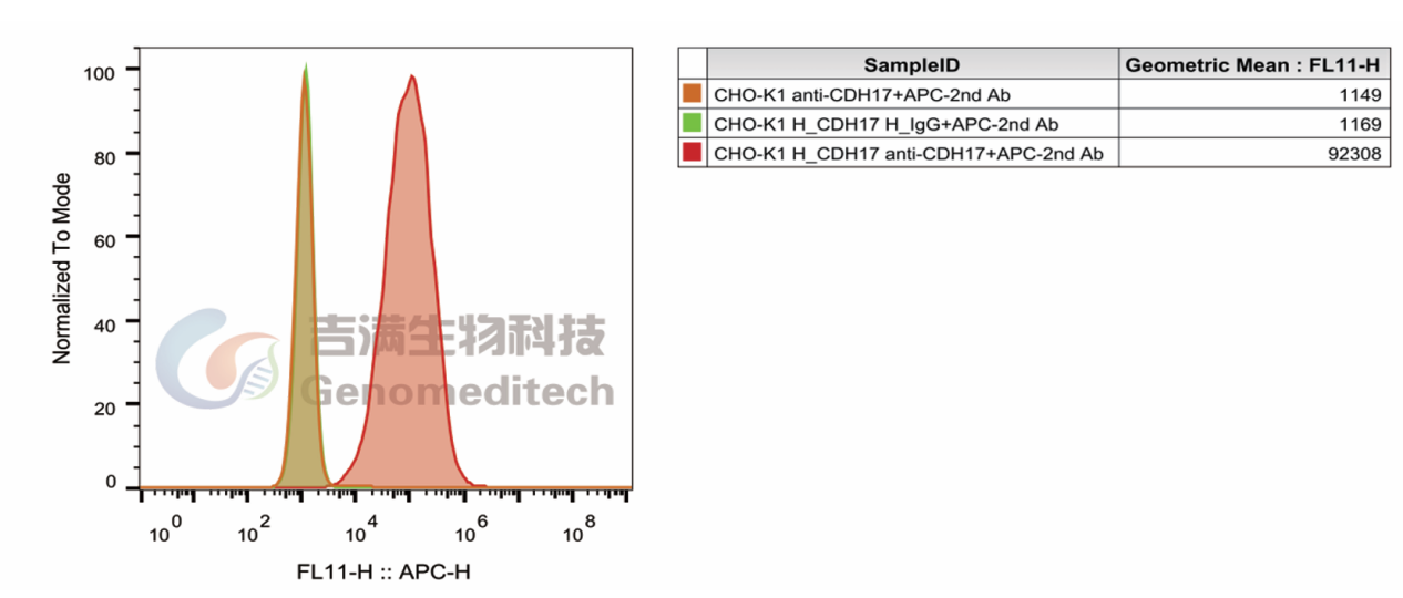
H_CDH17 CHO-K1 Cell Line (Catalog # GM-C25980) was stained with Anti-CDH17 hIgG1 Antibody (Catalog # GM-52672AB) or isotype control antibody, followed by anti-Human IgG APC-conjugated Secondary Antibody.
H_CDH17 CHO-K1 Cell Line, Anti-H_CDH17 hIgG1 Antibody(BI-905711)(Cat.GM-52672AB), Fold:79.0 |
|
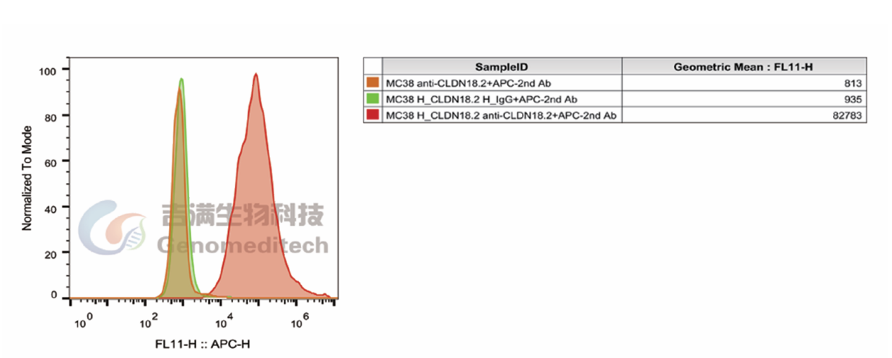
H_CLDN18(isoform2) eGFP 293 Cell Line (Catalog # GM-C02120) was stained with Anti CLDN18.2 hIgG1 Antibody(Zolbetuximab) (Catalog # GM-34137AB) or isotype control antibody, followed by anti-Human IgG APC-conjugated Secondary Antibody. |

Tumor immunotherapy has become an indispensable pillar of modern cancer treatment. The success of these therapies largely depends on the recognition of tumor antigens by T cells and the subsequent activation of the immune response.
Tumor antigens are generally divided into two categories: Tumor-Specific Antigens (TSAs) and Tumor-Associated Antigens (TAAs). TSAs are exclusively expressed on tumor cells and are typically absent or expressed at extremely low levels in normal cells, limiting their widespread clinical application. Conversely, TAAs usually arise from gene amplification, aberrant genetic regulation, or post-translational modifications. They exhibit high sharing and prevalence across the same tumor types among different patients.
TAAs are typically overexpressed on the surface of tumor cells while showing trace or selective expression in normal host tissues. When targeting specific malignancies, TAAs serve as highly promising therapeutic targets. They are now widely applied in the treatment of various tumors and play a critical role in the research and development of human oncology drugs. Antibodies targeting TAAs can not only mediate tumor cell killing through effector mechanisms such as ADCC (Antibody-Dependent Cellular Cytotoxicity) and CDC (Complement-Dependent Cytotoxicity), but can also serve as diagnostic biomarkers or be utilized to enhance the tumor-targeting specificity of traditional therapies.
Characteristics of TAAs
- Expressed not only in tumor cells but also partially present in normal host cells.
- Primarily driven by gene amplification, aberrant genetic regulation, or post-translational modifications, leading to upregulated expression in tumor cells.
- Exhibiting significantly higher expression levels in tumor cells compared to normal cells.
- Serving as diagnostic biomarkers or immunotherapy targets; they are critical targets for oncology immunotherapy strategies such as monoclonal antibodies, ADCs (Antibody-Drug Conjugates), and CAR-T (Chimeric Antigen Receptor T-cell) therapies.
We provide:
To support the research and development of tumor immunotherapy drugs, Genomeditech stays at the forefront of global biomedical R&D, meticulously building a comprehensive product library of TAA-related tool cell lines.
We currently offer 350+ off-the-shelf TAA-related products, covering 80+ target genes. These products faithfully recapitulate the presentation and native conformation of antigens on the cell surface, accelerating your drug screening and efficacy evaluation workflows.
|
Stable Cell Lines Based on HEK293 or CHO-K1 Backgrounds These cell lines, which stably express specific antigens, are primarily used for antibody-specific binding assays, affinity testing, and in vitro functional screening against relevant targets (e.g., CLDN18.2, AMHR2, CD123). |
Luciferase-Labeled Stable Cell Lines Based on Tumor Backgrounds These lines can be inoculated into mice to form solid tumors for the construction of CDX mouse models. Combined with in vivo optical imaging technology, they enable real-time, dynamic monitoring of tumor growth and metastasis in vivo. Widely utilized in in vivo efficacy evaluation, CAR-T cell therapy assessment, and the validation of other anti-tumor therapeutics. |
| Product No. | Product Name | Gene | Pre-order/order |
|
H_CDH17 CHO-K1 Cell Line (Catalog # GM-C25980) was stained with Anti-CDH17 hIgG1 Antibody (Catalog # GM-52672AB) or isotype control antibody, followed by anti-Human IgG APC-conjugated Secondary Antibody.
H_CDH17 CHO-K1 Cell Line, Anti-H_CDH17 hIgG1 Antibody(BI-905711)(Cat.GM-52672AB), Fold:79.0 |
|
H_CLDN18(isoform2) eGFP 293 Cell Line (Catalog # GM-C02120) was stained with Anti CLDN18.2 hIgG1 Antibody(Zolbetuximab) (Catalog # GM-34137AB) or isotype control antibody, followed by anti-Human IgG APC-conjugated Secondary Antibody. |