

NK cells, typically identified in humans as CD3-CD56+ cells, in several mouse strains as CD3-NK1.1+, comprise approximately 5-15% of circulating lymphocytes. Besides peripheral blood, NK cells are also found in non-lymphoid tissues such as the liver, uterus, adipose tissue, and intestines. In humans, NK cells can be divided into two subsets based on CD56 expression levels, CD56dim and CD56bright, reflecting cytotoxic and helper cell functions respectively.
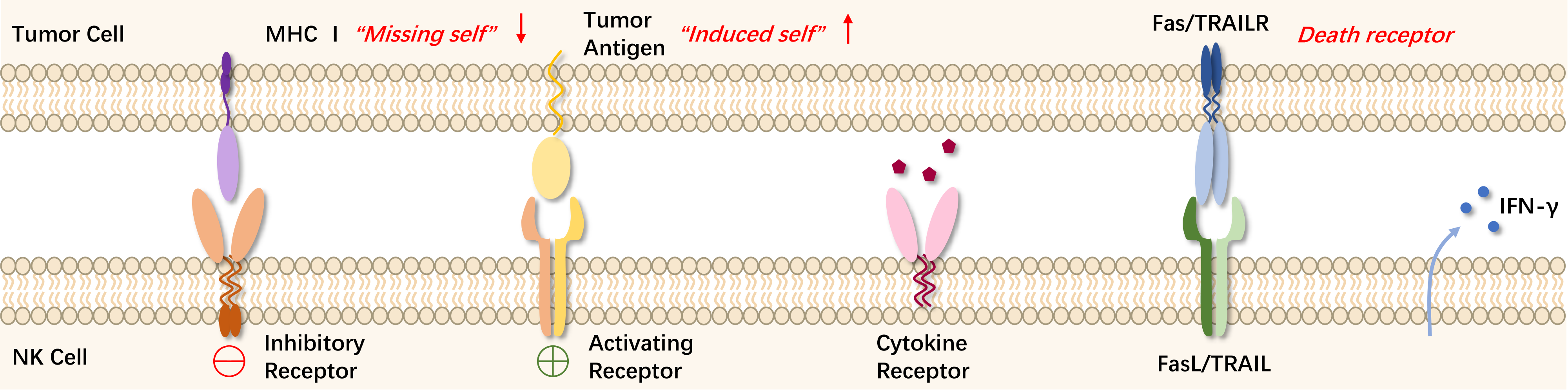
The killer cell immunoglobulin-like receptors (KIRs) expressed on the surface of NK cells can recognize major histocompatibility complex I (MHC I), which is widely expressed on normal cells and provides inhibitory signals to protect healthy cells from NK cell attacks. However, in cells experiencing viral infection or malignant transformation, MHC I expression is reduced or lost, allowing NK cells to escape KIR-induced inhibition and exert their cytolytic function. Aside from the "missing self" mechanism, NK cell-mediated immune surveillance also introduces the concept of "induced self". Activation receptors on NK cells include natural cytotoxicity receptors (NCR) and natural killer group 2D (NKG2D), which can recognize ligands on stressed cells and transmit activation signals. The balance between inhibitory and activating receptors determines the functional state of NK cells.