Where Discovery Begins
Jurkat CD3-BsAb Reporter Cell Line
Cat. No.
GM-C17940
Size
2 vial
Quote
Description
Data display
Specifications
Materials
Related products
Cell Culture
Description
Bispecific antibodies (BsAb) are an emerging cancer treatment strategy. Unlike traditional monoclonal antibodies, which use two Fab arms to recognize the same antigen, bispecific antibodies can recognize different antigens on each of their two binding arms. Among various bispecific antibodies, T Cell Engagers (TCEs) are particularly noteworthy because they can effectively enable T cells to recognize and kill tumor cells.

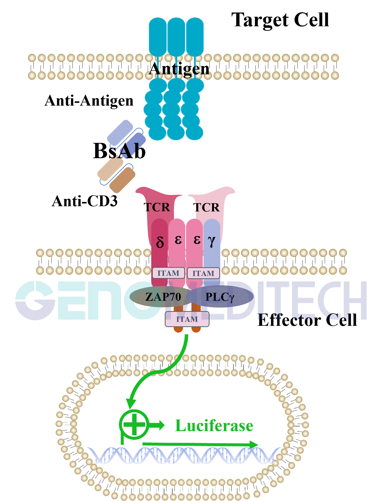
Jurkat CD3-BsAb Reporter Cell Line is a clonal stable Jurkat cell line with signal-dependent expression of a luciferase reporter gene constructed using lentiviral technology, and it endogenously expresses TCR-CD3 complex. T Cell Engager bispecific antibodies bridge T cells and tumor cells by binding to the TCR-CD3 complex on T cells and tumor-associated antigens on tumor cells, thereby activating the TCR signaling pathway. The bioactivity of T Cell Engager bispecific antibodies can be quantified by the production of luciferase, as TCR activation transmits signals to the cell nucleus, driving luciferase expression. Therefore, this cell line can be used as a tool for research on antibody drugs targeting T Cell Engagers.
Bispecific antibodies (BsAb) are an emerging cancer treatment strategy. Unlike traditional monoclonal antibodies, which use two Fab arms to recognize the same antigen, bispecific antibodies can recognize different antigens on each of their two binding arms. Among various bispecific antibodies, T Cell Engagers (TCEs) are particularly noteworthy because they can effectively enable T cells to recognize and kill tumor cells.

Jurkat CD3-BsAb Reporter Cell Line is a clonal stable Jurkat cell line with signal-dependent expression of a luciferase reporter gene constructed using lentiviral technology, and it endogenously expresses TCR-CD3 complex. T Cell Engager bispecific antibodies bridge T cells and tumor cells by binding to the TCR-CD3 complex on T cells and tumor-associated antigens on tumor cells, thereby activating the TCR signaling pathway. The bioactivity of T Cell Engager bispecific antibodies can be quantified by the production of luciferase, as TCR activation transmits signals to the cell nucleus, driving luciferase expression. Therefore, this cell line can be used as a tool for research on antibody drugs targeting T Cell Engagers.
Data display
Applications
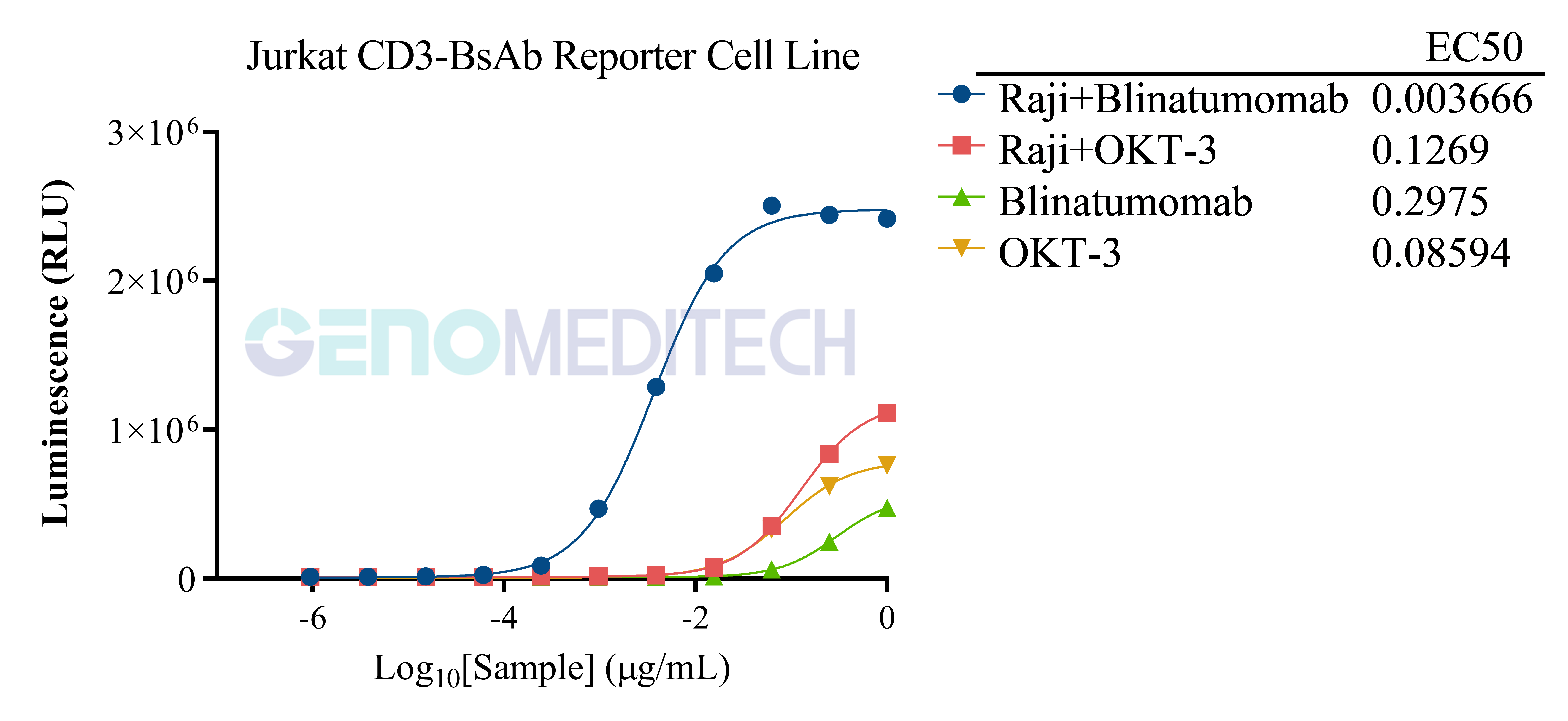
Response to Blinatumomab and OKT-3 (muromonab). Serial dilutions of the Anti-CD3-CD19 Bispecific Antibody(Blinatumomab) (Cat. GM-79712AB) or Anti-CD3 epsilon hIgG1 Antibody [OKT-3 (muromonab)] (Cat. GM-51478AB) were added to 1E5 cells/well of Raji cell line (Cat. GM-C19100), while the control group did not include Raji cells, and 1E5 cells/well of the Jurkat CD3-BsAb Reporter Cell Line (Cat. GM-C17940) for 7 hours. Firefly luciferase activity is then measured using the Luciferase Reporter Assay Kit (Genomeditech). Data are shown by drug mass concentration.
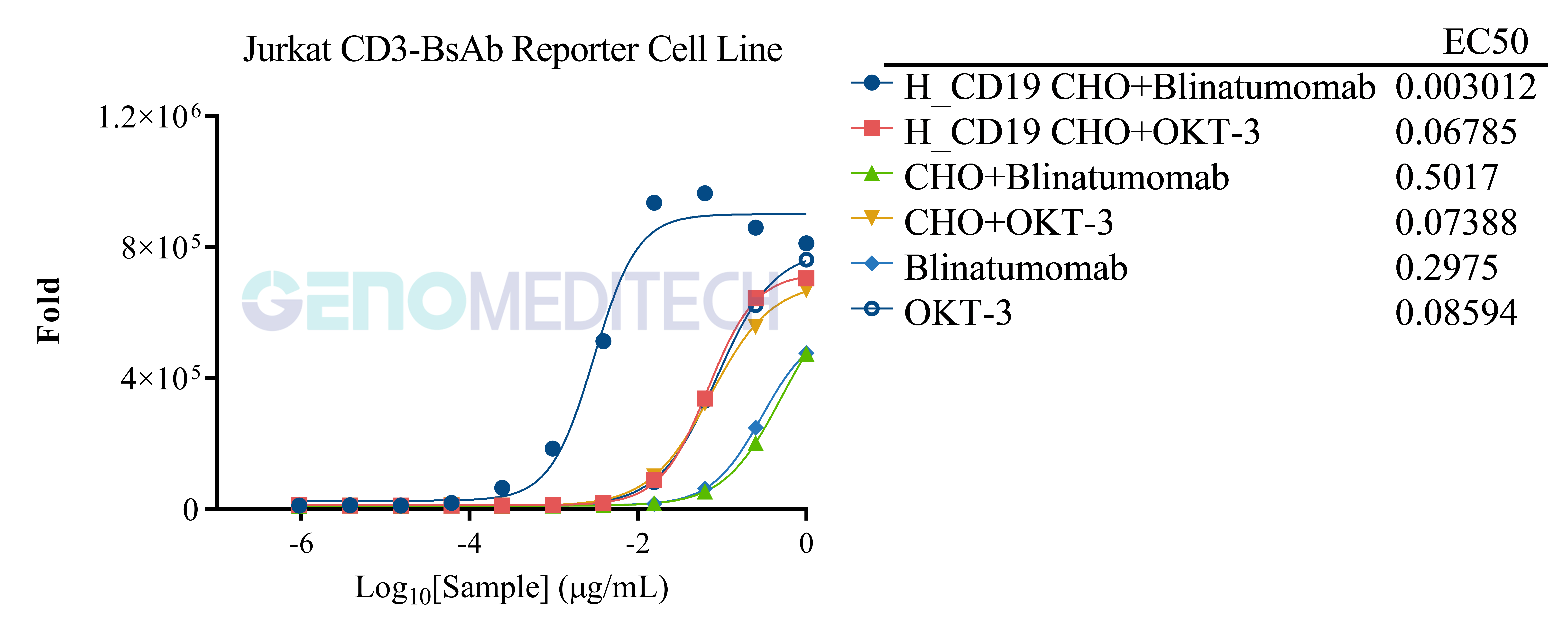
Response to Blinatumomab and OKT-3 (muromonab). Serial dilutions of the Anti-CD3-CD19 Bispecific Antibody(Blinatumomab) (Cat. GM-79712AB) or Anti-CD3 epsilon hIgG1 Antibody [OKT-3 (muromonab)] (Cat. GM-51478AB) were added to 1E4 cells/well of H_CD19 CHO-K1 Cell line (Cat. GM-C19025), while the control groups included CHO-K1 or did not include CHO-K1, and 1E5 cells/well of the Jurkat CD3-BsAb Reporter Cell Line (Cat. GM-C17940) for 7 hours. Firefly luciferase activity is then measured using the Luciferase Reporter Assay Kit (Genomeditech). Data are shown by drug mass concentration.
Expression
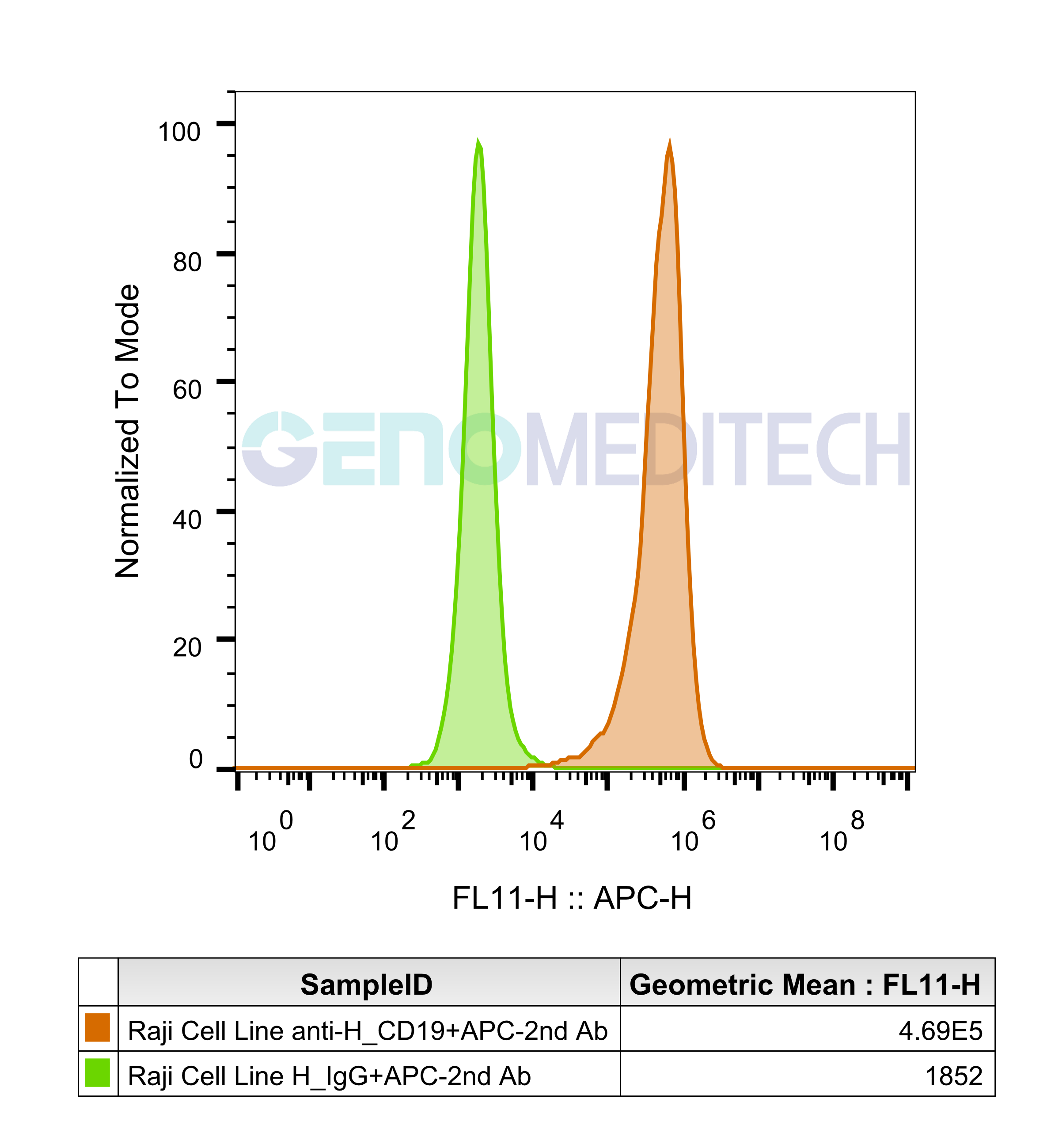
Raji Cell Line was determined by flow cytometry using Anti-H_CD19 hIgG1/hIgG2 Antibody (Cat. GM-28777AB).
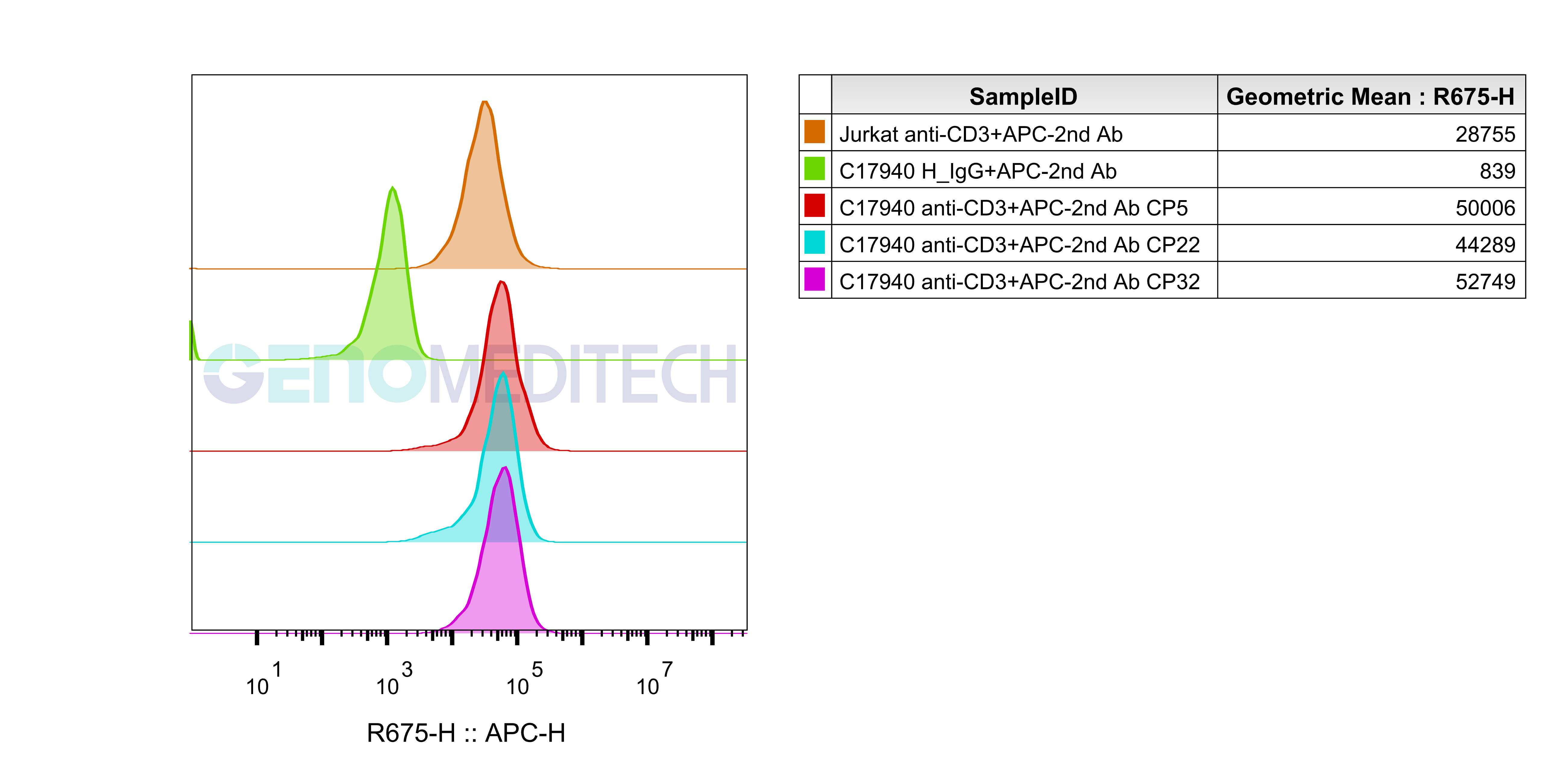
Jurkat CD3-BsAb Reporter Cell Line (Cat. GM-C17940) was determined by flow cytometry using Anti-CD3 hIgG1 Antibody(CH2527) (Cat. GM-33037AB).
Stability
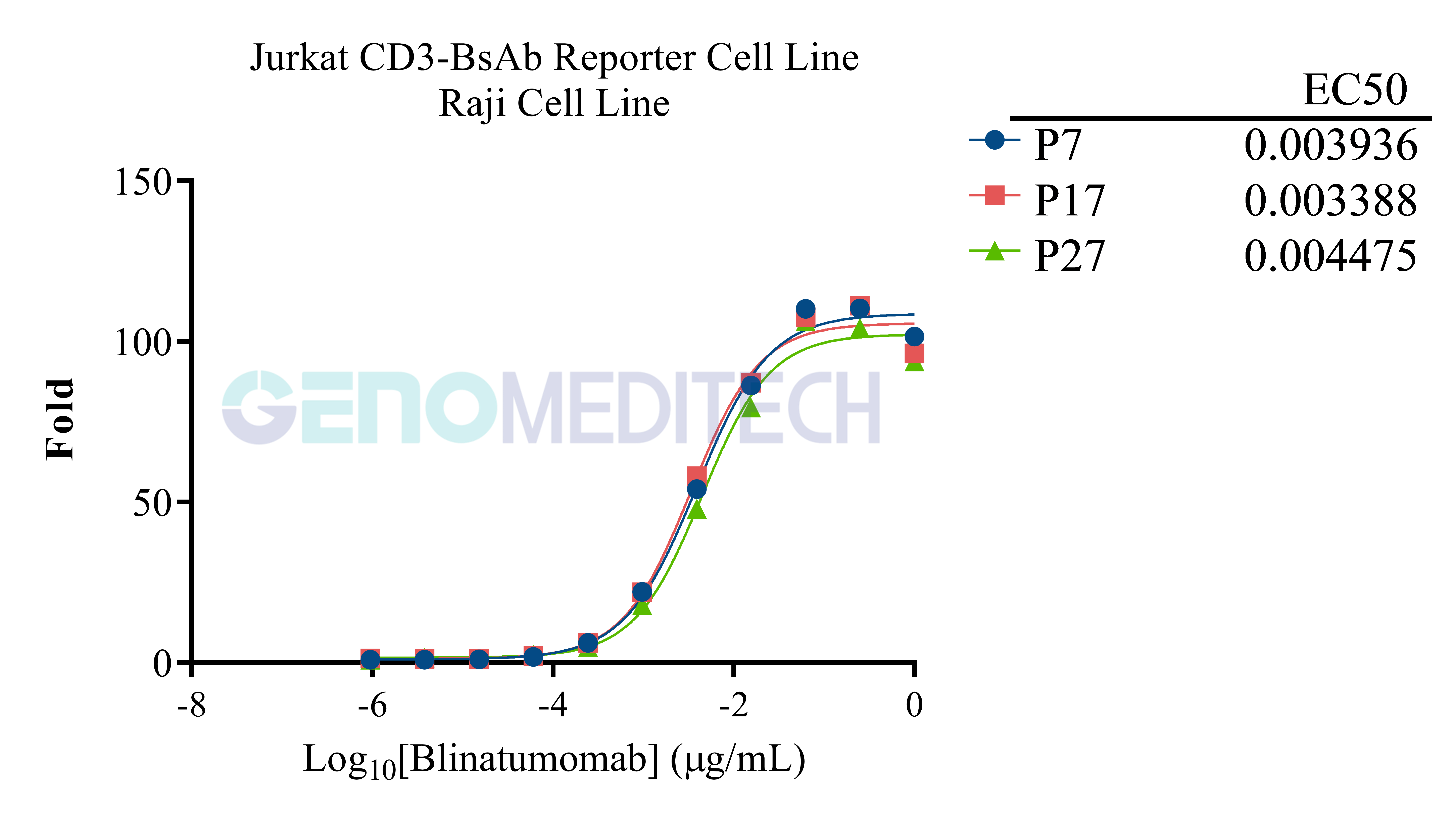
The passage stability of response to Anti-CD3-CD19 Bispecific Antibody. The passage 7, 17, and 27 of Jurkat CD3-BsAb Reporter Cell Line (Cat. GM-C17940) at a concentration of 1E5 cells/well was co-cultured with Raji cells (Cat. GM-C19100) that express CD19 endogenously at a concentration of 1E5 cells/well, in the presence of serial dilutions of the Anti-CD3-CD19 Bispecific Antibody (Cat. GM-79712AB) in assay buffer (RPMI 1640+1% FBS+1% P.S) for 7 hours (96-well format). The firefly luciferase activity was measured using the Luciferase Reporter Assay Kit (Genomeditech). Data are shown by drug mass concentration.
Specifications
Cat. NoGM-C17940
ProductJurkat CD3-BsAb Reporter Cell Line
Product Format1 vial of frozen cells
Quantity5E6 Cells per vial,1 mL
Storage ConditionsLiquid nitrogen immediately upon receipt
Recovery MediumRPMI 1640+10% FBS+1% P.S
Growth mediumRPMI 1640+10% FBS+1% P.S+3.5 μg/mL Blasticidin
NoteNone
Freezing Medium90% FBS+10% DMSO
Growth propertiesSuspension
Growth Conditions37°C, 5% CO₂
Safety considerationsBiosafety Level 2
Mycoplasma TestingThe cell line has been screened to confirm the absence of Mycoplasma species.
Cat. NoGM-C17940
ProductJurkat CD3-BsAb Reporter Cell Line
Product Format1 vial of frozen cells
Quantity5E6 Cells per vial,1 mL
Storage ConditionsLiquid nitrogen immediately upon receipt
Recovery MediumRPMI 1640+10% FBS+1% P.S
Growth mediumRPMI 1640+10% FBS+1% P.S+3.5 μg/mL Blasticidin
NoteNone
Freezing Medium90% FBS+10% DMSO
Growth propertiesSuspension
Growth Conditions37°C, 5% CO₂
Safety considerationsBiosafety Level 2
Mycoplasma TestingThe cell line has been screened to confirm the absence of Mycoplasma species.
Materials
ReagentOrdering Information
RPMI 1640gibco/C11875500BT
Fetal Bovine SerumExCell/FSP500
Pen/StrepThermo/15140-122
BlasticidinGenomeditech/GM-040404
Raji Cell LineGenomeditech/GM-C19100
H_CD19 CHO-K1 Cell lineGenomeditech/GM-C19025
Anti-CD3-CD19 Bispecific AntibodyGenomeditech/GM-79712AB
Anti-CD3 epsilon hIgG1 AntibodyGenomeditech/GM-51478AB
Anti-CD3 hIgG1 Antibody(CH2527)Genomeditech/GM-33037AB
Anti-H_CD19 hIgG1/hIgG2 AntibodyGenomeditech/GM-28777AB
GMOne-Step 2.0 Luciferase Reporter Gene Assay KitGenomeditech/GM-040513
96 well round cell culture platePakGent/CL-PT096
96-well White Opaque PlateThermo/236108
96-well U-bottom PlateSaining/1014010
Countess 3 Automated Cell CounterThermoFisher/A49891
SpectraMax® L Microplate LuminometerMoleculardevices/SpectraMax L
ReagentOrdering Information
RPMI 1640gibco/C11875500BT
Fetal Bovine SerumExCell/FSP500
Pen/StrepThermo/15140-122
BlasticidinGenomeditech/GM-040404
Raji Cell LineGenomeditech/GM-C19100
H_CD19 CHO-K1 Cell lineGenomeditech/GM-C19025
Anti-CD3-CD19 Bispecific AntibodyGenomeditech/GM-79712AB
Anti-CD3 epsilon hIgG1 AntibodyGenomeditech/GM-51478AB
Anti-CD3 hIgG1 Antibody(CH2527)Genomeditech/GM-33037AB
Anti-H_CD19 hIgG1/hIgG2 AntibodyGenomeditech/GM-28777AB
GMOne-Step 2.0 Luciferase Reporter Gene Assay KitGenomeditech/GM-040513
96 well round cell culture platePakGent/CL-PT096
96-well White Opaque PlateThermo/236108
96-well U-bottom PlateSaining/1014010
Countess 3 Automated Cell CounterThermoFisher/A49891
SpectraMax® L Microplate LuminometerMoleculardevices/SpectraMax L
Related products
CD28
H_CD28 Reporter Jurkat Cell LineCynomolgus_CD28 CHO-K1 Cell LineH_CD28 CHO-K1 Cell Line
H_CD28 HEK-293 Cell LineH_CD28 KO Jurkat Cell Line
Anti-CD28 hIgG4 AntibodyAnti-H_CD28 hIgG4 AntibodyAnti-mouse CD28 Syrian Hamster IgG2 Antibody(37. 51)
Human CD28 Protein; His Tag

CD19
H_CD19 KO Raji Cell LineCynomolgus_CD19 CHO-K1 Cell LineCynomolgus_CD19 HEK-293 Cell Line
H_CD19 CHO-K1 Cell lineH_CD19 HEK-293 Cell LineMouse_CD19 CHO-K1 Cell Line
Anti-CD19 hIgG1 Reference AntibodyAnti-CD19×CD3 hIgG1 AntibodyAnti-H_CD19 hIgG1/hIgG2 Antibody
CD3
Cynomolgus_CD3D CD3E Reporter Jurkat(CD3DE KO) Cell LineCynomolgus_CD3 HEK-293 Cell LineCynomolgus_CD3E(Membrane Bound ECD) CHO-K1 Cell Line
H_CD3 CHO-K1 Cell LineH_CD3 HEK-293 Cell LineH_CD3(TCR V2) CHO-K1 Cell Line
H_CD3(TCR V2) HEK-293 Cell LineH_CD3(TCRαβ delta V) HEK-293 Cell LineH_CD3D CD3E KO Jurkat Cell Line
H_CD3E KO Jurkat Cell LineH_CD3E(Membrane Bound ECD) CHO-K1 Cell LineMouse_CD3 HEK-293 Cell Line
Anti-CD19×CD3 hIgG1 AntibodyAnti-CD3 epsilon hIgG1 AntibodyAnti-CD3 hIgG1 Antibody
Anti-CD3×CD20 hIgG1 Bispecific Antibody Anti-CD3×FCRL5 hIgG1 Bispecific AntibodyAnti-CD3E×BCMA hIgG4 Reference Antibody
Anti-CD3E×DLL3 hIgG1 Bispecific AntibodyAnti-CD3E×MUC17 hIgG1 Bispecific AntibodyAnti-mouse CD3ε mIgG2a Antibody
Cynomolgus CD3E Protein; hFc TagHuman CD3E Protein; hFc Tag
CD2
H_CD2 KO Jurkat(CD3-) Cell LineCD3-CD2-tsAb Reporter Jurkat(CD58 KO) Cell LineCynomolgus_CD2 CHO-K1 Cell Line
H_CD2 CHO-K1 Cell LineH_CD2 HEK-293 Cell LineH_CD2 KO Jurkat(CD3+) Cell Line
Anti-CD19×CD3 hIgG1 AntibodyAnti-CD2 hIgG1 Antibody
Human CD2 Protein; hFc TagHuman CD2 Protein; His Tag
Cell Culture

Cell Recovery

Recovery Medium: RPMI 1640+10% FBS+1% P.S

To insure the highest level of viability, thaw the vial and initiate the culture as soon as possible upon receipt. If upon arrival, continued storage of the frozen culture is necessary, it should be stored in liquid nitrogen vapor phase and not at -70°C. Storage at -70°C will result in loss of viability.

a)       Thaw the vial by gentle agitation in a 37°C water bath. To reduce the possibility of contamination, keep the O-ring and cap out of the water. Thawing should be rapid (approximately 2 - 3 minutes).

b)      Remove the vial from the water bath as soon as the contents are thawed, and decontaminate by dipping in or spraying with 70% ethanol. All of the operations from this point on should be carried out under strict aseptic conditions.

c)       Transfer the vial contents to a centrifuge tube containing 5.0 mL complete culture medium. And spin at approximately 176 x g for 5 minutes. Discard supernatant.

d)      Resuspend cell pellet with the recommended complete medium. And dispense the suspension into 1 - 2 T-25 culture flasks.

e)       Incubate the culture at 37°C in a suitable incubator. A 5% CO₂ in air atmosphere is recommended if using the medium described on this product sheet.

Cell Freezing

Freezing Medium: 90% FBS+10% DMSO

a)         Centrifuge at 176 x g for 3 minutes to collect cells.

b)         Resuspend the cells in pre-cooled freezing medium and adjust the cell density to 5E6 cells/mL.

c)         Aliquot 1 mL into each vial.

d)         Place the vial in a controlled-rate freezing container and store at -80°C for at least 1 day, then transfer to liquid nitrogen as soon as possible.

Cell passage

Growth medium: RPMI 1640+10% FBS+1% P.S+3.5 μg/mL Blasticidin

Approximately 48-72 hours after the initial thawing, the cells can be passaged for the first time. After this initial passage, the culture medium can be adjusted to growth medium supplemented with antibiotics. If cells are not passaged within 48 hours, it is recommended to add some fresh recovery medium and place the flask horizontally.

a)         When the cell density reaches 1.5 - 2E6 cells/mL, subculture the cells. Do not allow the cell density to exceed 2E6 cells/mL.

b)         It is recommended to use T-25 flasks for subculturing.

c)         These cells are suspension cells, and it is recommended to use the "half-medium change" method to maintain optimal cell conditions during passaging.

d)         During passaging, you can directly add fresh growth medium to the culture flask, gently pipette to resuspend the cells, and then transfer the cell suspension to a new T-25 flask for continued culture.

Subcultivation Ratio: Maintain cultures at a cell concentraion between 3E5 and 1E6 viable cells/mL.

Medium Renewal: Every 2 to 3 days

Notes

a)         These cells are sensitive to density, so please ensure that the cell density is maintained within an appropriate range during culture and subculturing.

b)         During the first passage, pay attention to the nutrient supply; if not subculturing, make sure to add fresh recovery medium every other day as needed.


Cell Recovery

Recovery Medium: RPMI 1640+10% FBS+1% P.S

To insure the highest level of viability, thaw the vial and initiate the culture as soon as possible upon receipt. If upon arrival, continued storage of the frozen culture is necessary, it should be stored in liquid nitrogen vapor phase and not at -70°C. Storage at -70°C will result in loss of viability.

a)       Thaw the vial by gentle agitation in a 37°C water bath. To reduce the possibility of contamination, keep the O-ring and cap out of the water. Thawing should be rapid (approximately 2 - 3 minutes).

b)      Remove the vial from the water bath as soon as the contents are thawed, and decontaminate by dipping in or spraying with 70% ethanol. All of the operations from this point on should be carried out under strict aseptic conditions.

c)       Transfer the vial contents to a centrifuge tube containing 5.0 mL complete culture medium. And spin at approximately 176 x g for 5 minutes. Discard supernatant.

d)      Resuspend cell pellet with the recommended complete medium. And dispense the suspension into 1 - 2 T-25 culture flasks.

e)       Incubate the culture at 37°C in a suitable incubator. A 5% CO₂ in air atmosphere is recommended if using the medium described on this product sheet.

Cell Freezing

Freezing Medium: 90% FBS+10% DMSO

a)         Centrifuge at 176 x g for 3 minutes to collect cells.

b)         Resuspend the cells in pre-cooled freezing medium and adjust the cell density to 5E6 cells/mL.

c)         Aliquot 1 mL into each vial.

d)         Place the vial in a controlled-rate freezing container and store at -80°C for at least 1 day, then transfer to liquid nitrogen as soon as possible.

Cell passage

Growth medium: RPMI 1640+10% FBS+1% P.S+3.5 μg/mL Blasticidin

Approximately 48-72 hours after the initial thawing, the cells can be passaged for the first time. After this initial passage, the culture medium can be adjusted to growth medium supplemented with antibiotics. If cells are not passaged within 48 hours, it is recommended to add some fresh recovery medium and place the flask horizontally.

a)         When the cell density reaches 1.5 - 2E6 cells/mL, subculture the cells. Do not allow the cell density to exceed 2E6 cells/mL.

b)         It is recommended to use T-25 flasks for subculturing.

c)         These cells are suspension cells, and it is recommended to use the "half-medium change" method to maintain optimal cell conditions during passaging.

d)         During passaging, you can directly add fresh growth medium to the culture flask, gently pipette to resuspend the cells, and then transfer the cell suspension to a new T-25 flask for continued culture.

Subcultivation Ratio: Maintain cultures at a cell concentraion between 3E5 and 1E6 viable cells/mL.

Medium Renewal: Every 2 to 3 days

Notes

a)         These cells are sensitive to density, so please ensure that the cell density is maintained within an appropriate range during culture and subculturing.

b)         During the first passage, pay attention to the nutrient supply; if not subculturing, make sure to add fresh recovery medium every other day as needed.

For laboratory research use only. Direct human use, including taking orally and injection and clinical use are forbidden.
Jurkat CD3-BsAb Reporter Cell Line
Cat. No.
GM-C17940
Size
2 vial
Quote
Description
Data display
Specifications
Materials
Related products
Cell Culture
Description
Bispecific antibodies (BsAb) are an emerging cancer treatment strategy. Unlike traditional monoclonal antibodies, which use two Fab arms to recognize the same antigen, bispecific antibodies can recognize different antigens on each of their two binding arms. Among various bispecific antibodies, T Cell Engagers (TCEs) are particularly noteworthy because they can effectively enable T cells to recognize and kill tumor cells.

Jurkat CD3-BsAb Reporter Cell Line is a clonal stable Jurkat cell line with signal-dependent expression of a luciferase reporter gene constructed using lentiviral technology, and it endogenously expresses TCR-CD3 complex. T Cell Engager bispecific antibodies bridge T cells and tumor cells by binding to the TCR-CD3 complex on T cells and tumor-associated antigens on tumor cells, thereby activating the TCR signaling pathway. The bioactivity of T Cell Engager bispecific antibodies can be quantified by the production of luciferase, as TCR activation transmits signals to the cell nucleus, driving luciferase expression. Therefore, this cell line can be used as a tool for research on antibody drugs targeting T Cell Engagers.

Bispecific antibodies (BsAb) are an emerging cancer treatment strategy. Unlike traditional monoclonal antibodies, which use two Fab arms to recognize the same antigen, bispecific antibodies can recognize different antigens on each of their two binding arms. Among various bispecific antibodies, T Cell Engagers (TCEs) are particularly noteworthy because they can effectively enable T cells to recognize and kill tumor cells.

Jurkat CD3-BsAb Reporter Cell Line is a clonal stable Jurkat cell line with signal-dependent expression of a luciferase reporter gene constructed using lentiviral technology, and it endogenously expresses TCR-CD3 complex. T Cell Engager bispecific antibodies bridge T cells and tumor cells by binding to the TCR-CD3 complex on T cells and tumor-associated antigens on tumor cells, thereby activating the TCR signaling pathway. The bioactivity of T Cell Engager bispecific antibodies can be quantified by the production of luciferase, as TCR activation transmits signals to the cell nucleus, driving luciferase expression. Therefore, this cell line can be used as a tool for research on antibody drugs targeting T Cell Engagers.

Data display
Applications
Response to Blinatumomab and OKT-3 (muromonab). Serial dilutions of the Anti-CD3-CD19 Bispecific Antibody(Blinatumomab) (Cat. GM-79712AB) or Anti-CD3 epsilon hIgG1 Antibody [OKT-3 (muromonab)] (Cat. GM-51478AB) were added to 1E5 cells/well of Raji cell line (Cat. GM-C19100), while the control group did not include Raji cells, and 1E5 cells/well of the Jurkat CD3-BsAb Reporter Cell Line (Cat. GM-C17940) for 7 hours. Firefly luciferase activity is then measured using the Luciferase Reporter Assay Kit (Genomeditech). Data are shown by drug mass concentration.
Response to Blinatumomab and OKT-3 (muromonab). Serial dilutions of the Anti-CD3-CD19 Bispecific Antibody(Blinatumomab) (Cat. GM-79712AB) or Anti-CD3 epsilon hIgG1 Antibody [OKT-3 (muromonab)] (Cat. GM-51478AB) were added to 1E4 cells/well of H_CD19 CHO-K1 Cell line (Cat. GM-C19025), while the control groups included CHO-K1 or did not include CHO-K1, and 1E5 cells/well of the Jurkat CD3-BsAb Reporter Cell Line (Cat. GM-C17940) for 7 hours. Firefly luciferase activity is then measured using the Luciferase Reporter Assay Kit (Genomeditech). Data are shown by drug mass concentration.
Expression
Raji Cell Line was determined by flow cytometry using Anti-H_CD19 hIgG1/hIgG2 Antibody (Cat. GM-28777AB).
Jurkat CD3-BsAb Reporter Cell Line (Cat. GM-C17940) was determined by flow cytometry using Anti-CD3 hIgG1 Antibody(CH2527) (Cat. GM-33037AB).
Stability
The passage stability of response to Anti-CD3-CD19 Bispecific Antibody. The passage 7, 17, and 27 of Jurkat CD3-BsAb Reporter Cell Line (Cat. GM-C17940) at a concentration of 1E5 cells/well was co-cultured with Raji cells (Cat. GM-C19100) that express CD19 endogenously at a concentration of 1E5 cells/well, in the presence of serial dilutions of the Anti-CD3-CD19 Bispecific Antibody (Cat. GM-79712AB) in assay buffer (RPMI 1640+1% FBS+1% P.S) for 7 hours (96-well format). The firefly luciferase activity was measured using the Luciferase Reporter Assay Kit (Genomeditech). Data are shown by drug mass concentration.
Specifications
Cat. NoGM-C17940
ProductJurkat CD3-BsAb Reporter Cell Line
Product Format1 vial of frozen cells
Quantity5E6 Cells per vial,1 mL
Storage ConditionsLiquid nitrogen immediately upon receipt
Recovery MediumRPMI 1640+10% FBS+1% P.S
Growth mediumRPMI 1640+10% FBS+1% P.S+3.5 μg/mL Blasticidin
NoteNone
Freezing Medium90% FBS+10% DMSO
Growth propertiesSuspension
Growth Conditions37°C, 5% CO₂
Safety considerationsBiosafety Level 2
Mycoplasma TestingThe cell line has been screened to confirm the absence of Mycoplasma species.
Cat. NoGM-C17940
ProductJurkat CD3-BsAb Reporter Cell Line
Product Format1 vial of frozen cells
Quantity5E6 Cells per vial,1 mL
Storage ConditionsLiquid nitrogen immediately upon receipt
Recovery MediumRPMI 1640+10% FBS+1% P.S
Growth mediumRPMI 1640+10% FBS+1% P.S+3.5 μg/mL Blasticidin
NoteNone
Freezing Medium90% FBS+10% DMSO
Growth propertiesSuspension
Growth Conditions37°C, 5% CO₂
Safety considerationsBiosafety Level 2
Mycoplasma TestingThe cell line has been screened to confirm the absence of Mycoplasma species.
Materials
ReagentOrdering Information
RPMI 1640gibco/C11875500BT
Fetal Bovine SerumExCell/FSP500
Pen/StrepThermo/15140-122
BlasticidinGenomeditech/GM-040404
Raji Cell LineGenomeditech/GM-C19100
H_CD19 CHO-K1 Cell lineGenomeditech/GM-C19025
Anti-CD3-CD19 Bispecific AntibodyGenomeditech/GM-79712AB
Anti-CD3 epsilon hIgG1 AntibodyGenomeditech/GM-51478AB
Anti-CD3 hIgG1 Antibody(CH2527)Genomeditech/GM-33037AB
Anti-H_CD19 hIgG1/hIgG2 AntibodyGenomeditech/GM-28777AB
GMOne-Step 2.0 Luciferase Reporter Gene Assay KitGenomeditech/GM-040513
96 well round cell culture platePakGent/CL-PT096
96-well White Opaque PlateThermo/236108
96-well U-bottom PlateSaining/1014010
Countess 3 Automated Cell CounterThermoFisher/A49891
SpectraMax® L Microplate LuminometerMoleculardevices/SpectraMax L
ReagentOrdering Information
RPMI 1640gibco/C11875500BT
Fetal Bovine SerumExCell/FSP500
Pen/StrepThermo/15140-122
BlasticidinGenomeditech/GM-040404
Raji Cell LineGenomeditech/GM-C19100
H_CD19 CHO-K1 Cell lineGenomeditech/GM-C19025
Anti-CD3-CD19 Bispecific AntibodyGenomeditech/GM-79712AB
Anti-CD3 epsilon hIgG1 AntibodyGenomeditech/GM-51478AB
Anti-CD3 hIgG1 Antibody(CH2527)Genomeditech/GM-33037AB
Anti-H_CD19 hIgG1/hIgG2 AntibodyGenomeditech/GM-28777AB
GMOne-Step 2.0 Luciferase Reporter Gene Assay KitGenomeditech/GM-040513
96 well round cell culture platePakGent/CL-PT096
96-well White Opaque PlateThermo/236108
96-well U-bottom PlateSaining/1014010
Countess 3 Automated Cell CounterThermoFisher/A49891
SpectraMax® L Microplate LuminometerMoleculardevices/SpectraMax L
Related products
CD28
H_CD28 Reporter Jurkat Cell LineCynomolgus_CD28 CHO-K1 Cell LineH_CD28 CHO-K1 Cell Line
H_CD28 HEK-293 Cell LineH_CD28 KO Jurkat Cell Line
Anti-CD28 hIgG4 AntibodyAnti-H_CD28 hIgG4 AntibodyAnti-mouse CD28 Syrian Hamster IgG2 Antibody(37. 51)
Human CD28 Protein; His Tag

CD19
H_CD19 KO Raji Cell LineCynomolgus_CD19 CHO-K1 Cell LineCynomolgus_CD19 HEK-293 Cell Line
H_CD19 CHO-K1 Cell lineH_CD19 HEK-293 Cell LineMouse_CD19 CHO-K1 Cell Line
Anti-CD19 hIgG1 Reference AntibodyAnti-CD19×CD3 hIgG1 AntibodyAnti-H_CD19 hIgG1/hIgG2 Antibody
CD3
Cynomolgus_CD3D CD3E Reporter Jurkat(CD3DE KO) Cell LineCynomolgus_CD3 HEK-293 Cell LineCynomolgus_CD3E(Membrane Bound ECD) CHO-K1 Cell Line
H_CD3 CHO-K1 Cell LineH_CD3 HEK-293 Cell LineH_CD3(TCR V2) CHO-K1 Cell Line
H_CD3(TCR V2) HEK-293 Cell LineH_CD3(TCRαβ delta V) HEK-293 Cell LineH_CD3D CD3E KO Jurkat Cell Line
H_CD3E KO Jurkat Cell LineH_CD3E(Membrane Bound ECD) CHO-K1 Cell LineMouse_CD3 HEK-293 Cell Line
Anti-CD19×CD3 hIgG1 AntibodyAnti-CD3 epsilon hIgG1 AntibodyAnti-CD3 hIgG1 Antibody
Anti-CD3×CD20 hIgG1 Bispecific Antibody Anti-CD3×FCRL5 hIgG1 Bispecific AntibodyAnti-CD3E×BCMA hIgG4 Reference Antibody
Anti-CD3E×DLL3 hIgG1 Bispecific AntibodyAnti-CD3E×MUC17 hIgG1 Bispecific AntibodyAnti-mouse CD3ε mIgG2a Antibody
Cynomolgus CD3E Protein; hFc TagHuman CD3E Protein; hFc Tag
CD2
H_CD2 KO Jurkat(CD3-) Cell LineCD3-CD2-tsAb Reporter Jurkat(CD58 KO) Cell LineCynomolgus_CD2 CHO-K1 Cell Line
H_CD2 CHO-K1 Cell LineH_CD2 HEK-293 Cell LineH_CD2 KO Jurkat(CD3+) Cell Line
Anti-CD19×CD3 hIgG1 AntibodyAnti-CD2 hIgG1 Antibody
Human CD2 Protein; hFc TagHuman CD2 Protein; His Tag
Cell Culture

Cell Recovery

Recovery Medium: RPMI 1640+10% FBS+1% P.S

To insure the highest level of viability, thaw the vial and initiate the culture as soon as possible upon receipt. If upon arrival, continued storage of the frozen culture is necessary, it should be stored in liquid nitrogen vapor phase and not at -70°C. Storage at -70°C will result in loss of viability.

a)       Thaw the vial by gentle agitation in a 37°C water bath. To reduce the possibility of contamination, keep the O-ring and cap out of the water. Thawing should be rapid (approximately 2 - 3 minutes).

b)      Remove the vial from the water bath as soon as the contents are thawed, and decontaminate by dipping in or spraying with 70% ethanol. All of the operations from this point on should be carried out under strict aseptic conditions.

c)       Transfer the vial contents to a centrifuge tube containing 5.0 mL complete culture medium. And spin at approximately 176 x g for 5 minutes. Discard supernatant.

d)      Resuspend cell pellet with the recommended complete medium. And dispense the suspension into 1 - 2 T-25 culture flasks.

e)       Incubate the culture at 37°C in a suitable incubator. A 5% CO₂ in air atmosphere is recommended if using the medium described on this product sheet.

Cell Freezing

Freezing Medium: 90% FBS+10% DMSO

a)         Centrifuge at 176 x g for 3 minutes to collect cells.

b)         Resuspend the cells in pre-cooled freezing medium and adjust the cell density to 5E6 cells/mL.

c)         Aliquot 1 mL into each vial.

d)         Place the vial in a controlled-rate freezing container and store at -80°C for at least 1 day, then transfer to liquid nitrogen as soon as possible.

Cell passage

Growth medium: RPMI 1640+10% FBS+1% P.S+3.5 μg/mL Blasticidin

Approximately 48-72 hours after the initial thawing, the cells can be passaged for the first time. After this initial passage, the culture medium can be adjusted to growth medium supplemented with antibiotics. If cells are not passaged within 48 hours, it is recommended to add some fresh recovery medium and place the flask horizontally.

a)         When the cell density reaches 1.5 - 2E6 cells/mL, subculture the cells. Do not allow the cell density to exceed 2E6 cells/mL.

b)         It is recommended to use T-25 flasks for subculturing.

c)         These cells are suspension cells, and it is recommended to use the "half-medium change" method to maintain optimal cell conditions during passaging.

d)         During passaging, you can directly add fresh growth medium to the culture flask, gently pipette to resuspend the cells, and then transfer the cell suspension to a new T-25 flask for continued culture.

Subcultivation Ratio: Maintain cultures at a cell concentraion between 3E5 and 1E6 viable cells/mL.

Medium Renewal: Every 2 to 3 days

Notes

a)         These cells are sensitive to density, so please ensure that the cell density is maintained within an appropriate range during culture and subculturing.

b)         During the first passage, pay attention to the nutrient supply; if not subculturing, make sure to add fresh recovery medium every other day as needed.


Contact
Us
Message Inquiry
reset
submit
Message
Message Inquiry
reset
submit