Where Discovery Begins
H_CD25 CD122 CD132 Reporter Jurkat(hPD1 OE) Cell Line
Cat. No.
GM-C41979
Size
2 vial
Quote
Description
Data display
Specifications
Materials
Cell Culture
Related products
Description
IL-2, secreted by activated T cells, promotes effector and naïve T cell survival/proliferation but also activates Tregs. IL-2R exists as high-affinity trimers (CD25+CD122+CD132), intermediate-affinity dimers (CD122+CD132), or low-affinity CD25 monomers.
PD-1 is an immune checkpoint receptor on activated T, B, and myeloid cells. Upon binding PD-L1/L2, its ITIM/ITSM motifs recruit SHP2, suppressing TCR/CD28 signaling and reducing T cell proliferation, cytotoxicity, and cytokine secretion to maintain peripheral tolerance.

Anti-PD1-IL2v is a novel immunotherapeutic that combines an anti-PD-1 monoclonal antibody with an engineered IL-2 variant (IL2v) via fusion or conjugation.

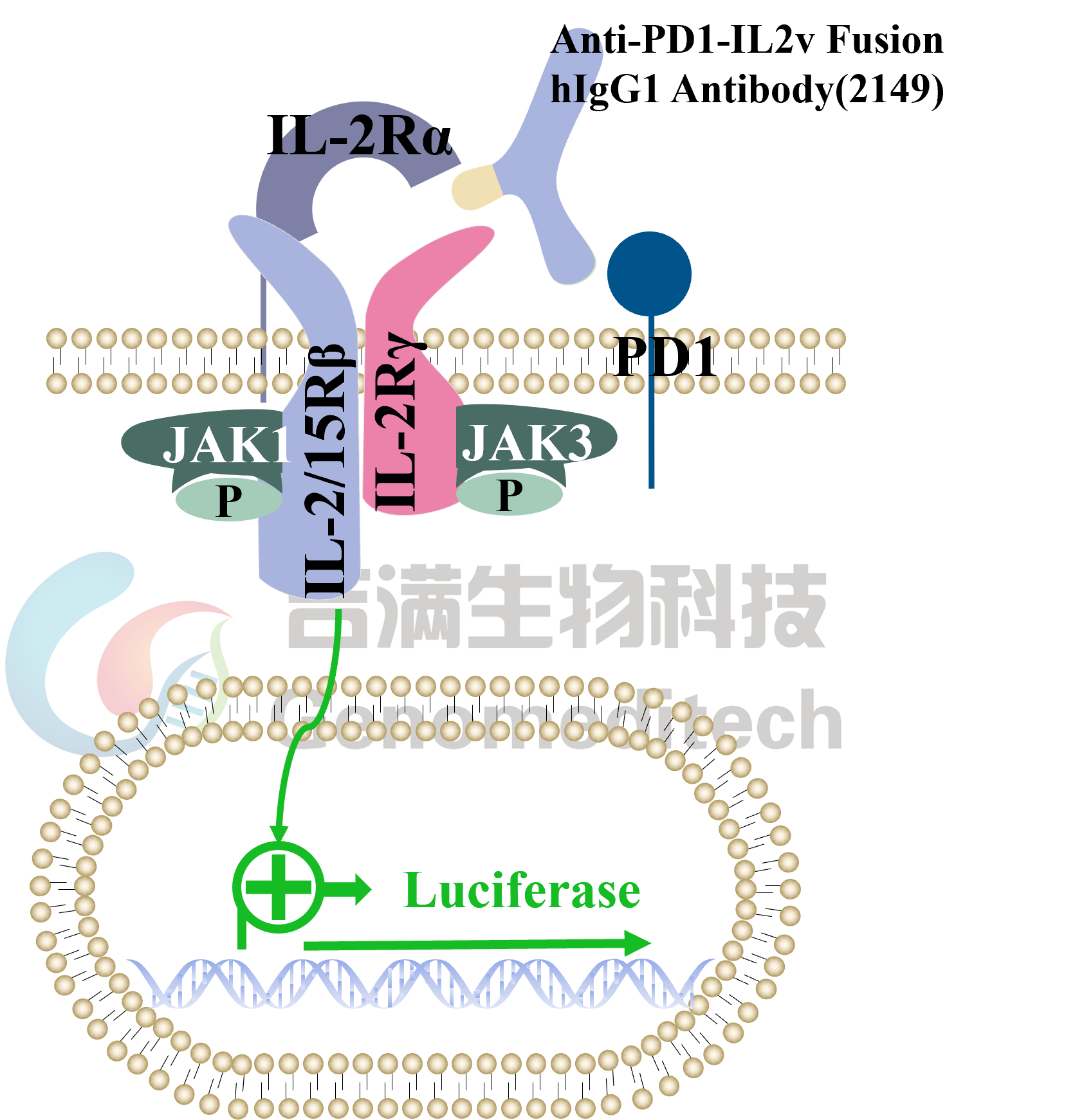
H_CD25 CD122 CD132 Reporter Jurkat(hPD1 OE) Cell Line is a stable clonal Jurkat cell line constructed using lentiviral technology, constitutive expression of the CD25,CD122,CD132 and PD1 genes, along with signal-dependent expression of a luciferase reporter gene. When the Anti-PD-1-IL-2v fusion protein binds to both PD-1 and IL-2R on the cell surface, it triggers IL-2R downstream signaling, leading to luciferase expression. The luciferase signal quantifies the level of pathway activation. Thus, this system allows for evaluating PD-1-dependent IL-2R activation enhancement and is suitable for in vitro testing of related drugs.
IL-2, secreted by activated T cells, promotes effector and naïve T cell survival/proliferation but also activates Tregs. IL-2R exists as high-affinity trimers (CD25+CD122+CD132), intermediate-affinity dimers (CD122+CD132), or low-affinity CD25 monomers.
PD-1 is an immune checkpoint receptor on activated T, B, and myeloid cells. Upon binding PD-L1/L2, its ITIM/ITSM motifs recruit SHP2, suppressing TCR/CD28 signaling and reducing T cell proliferation, cytotoxicity, and cytokine secretion to maintain peripheral tolerance.

Anti-PD1-IL2v is a novel immunotherapeutic that combines an anti-PD-1 monoclonal antibody with an engineered IL-2 variant (IL2v) via fusion or conjugation.

H_CD25 CD122 CD132 Reporter Jurkat(hPD1 OE) Cell Line is a stable clonal Jurkat cell line constructed using lentiviral technology, constitutive expression of the CD25,CD122,CD132 and PD1 genes, along with signal-dependent expression of a luciferase reporter gene. When the Anti-PD-1-IL-2v fusion protein binds to both PD-1 and IL-2R on the cell surface, it triggers IL-2R downstream signaling, leading to luciferase expression. The luciferase signal quantifies the level of pathway activation. Thus, this system allows for evaluating PD-1-dependent IL-2R activation enhancement and is suitable for in vitro testing of related drugs.
Data display
Applications
Response to Anti-PD1-IL2v Fusion hIgG1 Antibody(2149). The H_CD25 CD122 CD132 Reporter Jurkat(hPD1 OE) Cell Line (Cat. GM-C41979) at a concentration of 1E5 cells/well (96-well format) was stimulated with serial dilutions of Anti-PD1-IL2v (Cat. GM-88264AB) in assay buffer (RPMI 1640+1% FBS+1% P.S) for 16 hours. The firefly luciferase activity was measured using the Luciferase Reporter Assay Kit (Genomeditech). The maximum induction fold was approximately [10.5]. Data are shown by drug molar concentration.
Response to Recombinant Anti-PD1-IL2v Fusion hIgG1 Antibody(2149). H_CD25 CD122 CD132 Reporter Jurkat(hPD1 OE) Cell Line (Cat. GM-C41979) and H_CD25 CD122 CD132 Reporter Cell Line (Cat. GM-C29055) at a concentration of 1E5 cells/well (96-well format) were stimulated with serial dilutions of Anti-PD1-IL2v Fusion hIgG1 Antibody(2149) (Cat.GM-88264AB) in assay buffer (RPMI 1640 + 1% FBS + 1%P.S) for 16 hours. The firefly luciferase activity was measured using the Luciferase Reporter Assay Kit (Genomeditech). The maximum induction fold for the H_CD25 CD122 CD132 Reporter Jurkat(hPD1 OE) was approximately [10.5], and the H_CD25 CD122 CD132 Reporter validation was invalid/failed. Data are shown by drug molar concentration.
Signaling Pathway
Response to Recombinant Human IL-2. The H_CD25 CD122 CD132 Reporter Jurkat(hPD1 OE) Cell Line (Cat. GM-C41979) at a concentration of 1E5 cells/well (96-well format) was stimulated with serial dilutions of Recombinant Human IL-2 (Novoprotein/C013)in assay buffer (RPMI 1640+1% FBS+1% P.S) for 16 hours. The firefly luciferase activity was measured using the Luciferase Reporter Assay Kit (Genomeditech). The maximum induction fold was approximately [22.5]. Data are shown by drug molar concentration.
Specificity
Response to Recombinant Human IL-2. The H_CD25 CD122 CD132 Reporter Cell Line (Cat. GM-C29055) at a concentration of 1E5 cells/well (96-well format) was stimulated with serial dilutions of Recombinant Human IL-2 (Novoprotein/C013)in assay buffer (RPMI 1640+1% FBS+1% P.S) for 16 hours. The firefly luciferase activity was measured using the Luciferase Reporter Assay Kit (Genomeditech). The maximum induction fold was approximately [6.7]. Data are shown by drug molar concentration.
Expression
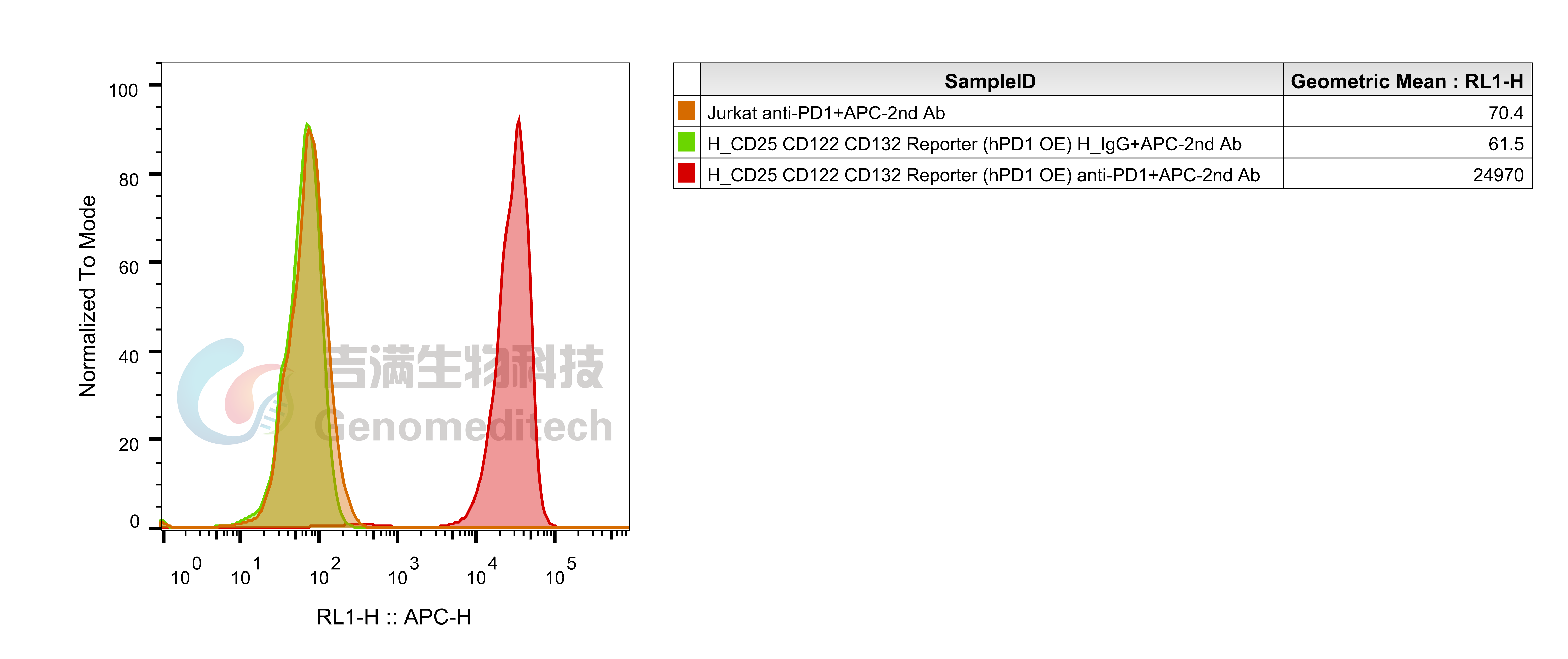
H_CD25 CD122 CD132 Reporter Jurkat(hPD1 OE) Cell Line (Cat. GM-C41979) was determined by flow cytometry using Anti-PD1 hIgG4 Reference Antibody (Pembio) (Cat. GM-87802MAB).
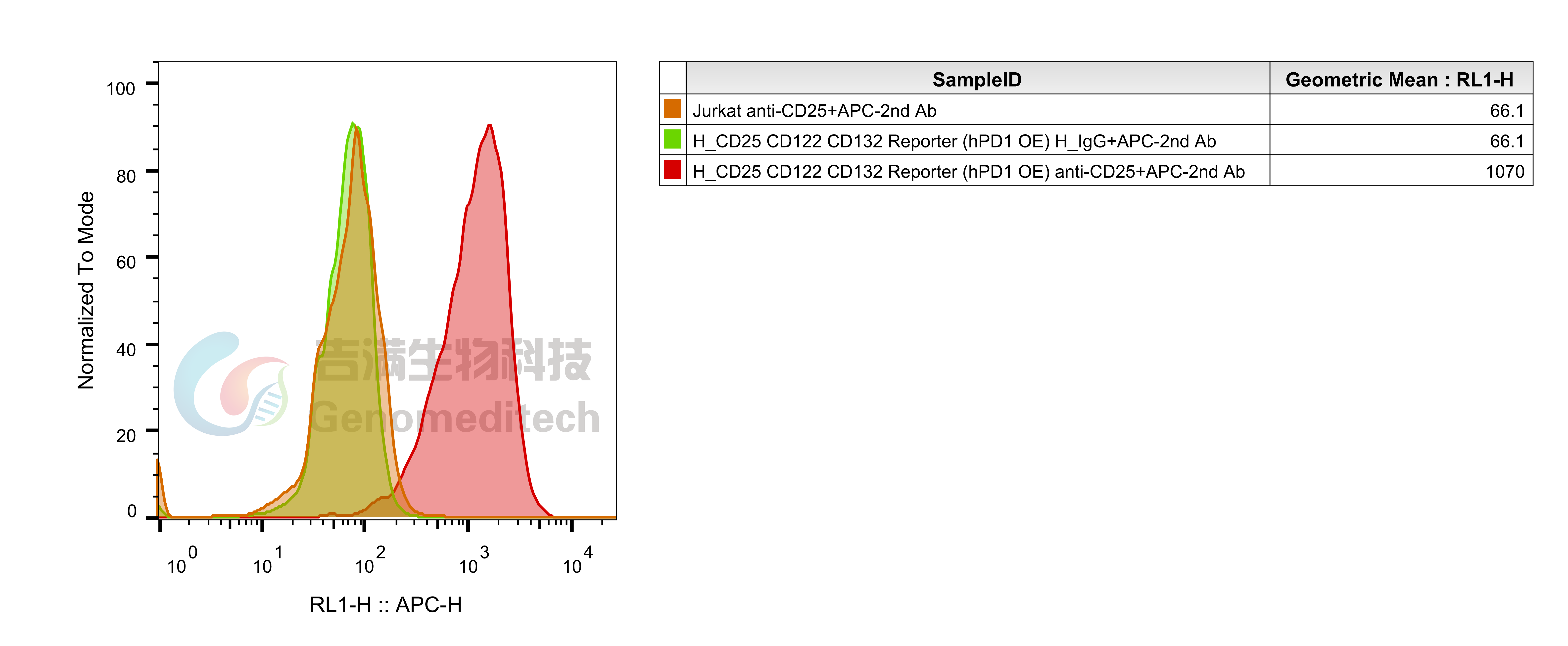
H_CD25 CD122 CD132 Reporter Jurkat(hPD1 OE) Cell Line (Cat. GM-C41979) was determined by flow cytometry using Anti-CD25 hIgG1 Antibody (Cat. GM-52329AB).
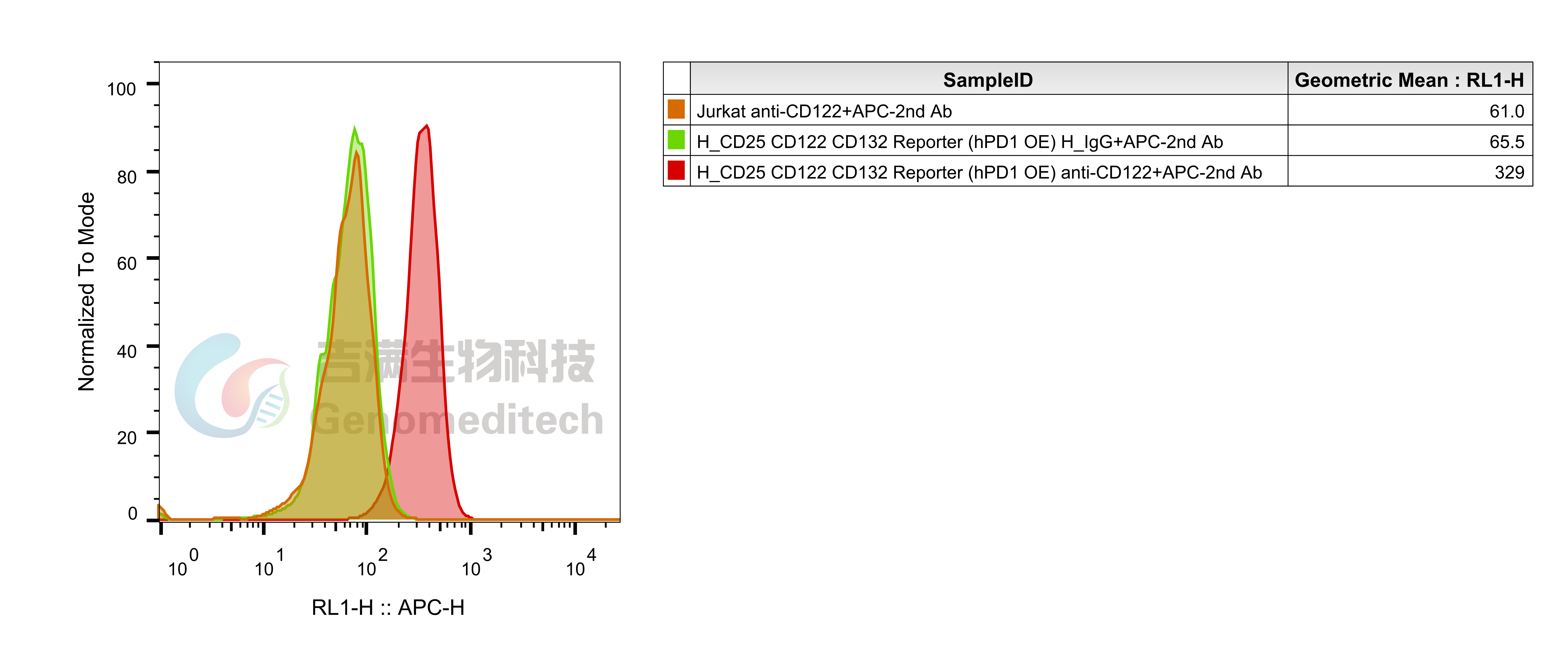
H_CD25 CD122 CD132 Reporter Jurkat(hPD1 OE) Cell Line (Cat. GM-C41979) was determined by flow cytometry using Anti-CD122 hIgG1 Antibody(HuABC-2) (Cat. GM-52319AB).
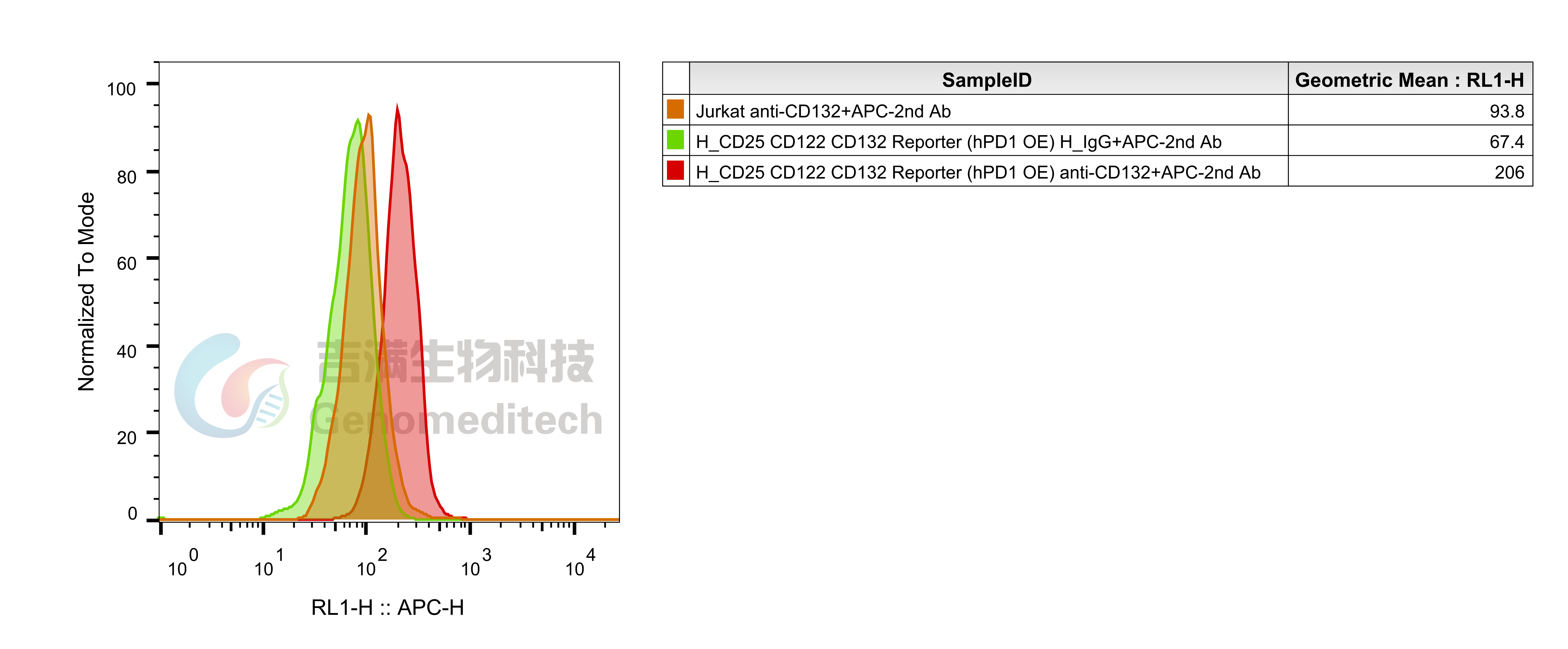
H_CD25 CD122 CD132 Reporter Jurkat(hPD1 OE) Cell Line (Cat. GM-C41979) was determined by flow cytometry using Anti-CD132(IL2RG) hIgG4 Antibody(REGN7257) (Cat. GM-52334AB).
Specifications
Cat. No GM-C41979
Product H_CD25 CD122 CD132 Reporter Jurkat(hPD1 OE) Cell Line
Product Format 1 vial of frozen cells
Quantity 5E6 Cells per vial,1 mL
Storage Conditions Liquid nitrogen immediately upon receipt
Recovery Medium RPMI 1640+10% FBS+1% P.S
Growth medium RPMI 1640+10% FBS+1% P.S+3.5 μg/mL Blasticidin+400 μg/mL Bleomycin+400 μg/mL G418+200 μg/mL Hygromycin+0.75 μg/mL Puromycin
Note None
Freezing Medium 90% FBS+10% DMSO
Growth properties Suspension
Growth Conditions 37°C, 5% CO₂
Safety considerations Biosafety Level 2
Mycoplasma Testing The cell line has been screened to confirm the absence of Mycoplasma species.
Cat. No GM-C41979
Product H_CD25 CD122 CD132 Reporter Jurkat(hPD1 OE) Cell Line
Product Format 1 vial of frozen cells
Quantity 5E6 Cells per vial,1 mL
Storage Conditions Liquid nitrogen immediately upon receipt
Recovery Medium RPMI 1640+10% FBS+1% P.S
Growth medium RPMI 1640+10% FBS+1% P.S+3.5 μg/mL Blasticidin+400 μg/mL Bleomycin+400 μg/mL G418+200 μg/mL Hygromycin+0.75 μg/mL Puromycin
Note None
Freezing Medium 90% FBS+10% DMSO
Growth properties Suspension
Growth Conditions 37°C, 5% CO₂
Safety considerations Biosafety Level 2
Mycoplasma Testing The cell line has been screened to confirm the absence of Mycoplasma species.
Materials
Reagent Ordering Information
Fetal Bovine Serum ExCell/FSP500
Pen/Strep Thermo/15140-122
Blasticidin Genomeditech/GM-040404
G418 Genomeditech/GM-040402
Hygromycin Genomeditech/GM-040403
Puromycin Genomeditech/GM-040401
Bleomycin Genomeditech/GM-040407
Anti-PD1-IL2v Fusion hIgG1 Antibody(2149) Genomeditech/GM-88264AB
Anti-PD1 hIgG4 Reference Antibody (Pembio) Genomeditech/GM-87802MAB
Anti-CD25 hIgG1 Antibody Genomeditech/GM-52329AB
Anti-CD122 hIgG1 Antibody(HuABC-2) Genomeditech/GM-52319AB
Anti-CD132(IL2RG) hIgG4 Antibody(REGN7257) Genomeditech/GM-52334AB
GMOne-Step 2.0 Luciferase Reporter Gene Assay Kit Genomeditech/GM-040513
Reagent Ordering Information
Fetal Bovine Serum ExCell/FSP500
Pen/Strep Thermo/15140-122
Blasticidin Genomeditech/GM-040404
G418 Genomeditech/GM-040402
Hygromycin Genomeditech/GM-040403
Puromycin Genomeditech/GM-040401
Bleomycin Genomeditech/GM-040407
Anti-PD1-IL2v Fusion hIgG1 Antibody(2149) Genomeditech/GM-88264AB
Anti-PD1 hIgG4 Reference Antibody (Pembio) Genomeditech/GM-87802MAB
Anti-CD25 hIgG1 Antibody Genomeditech/GM-52329AB
Anti-CD122 hIgG1 Antibody(HuABC-2) Genomeditech/GM-52319AB
Anti-CD132(IL2RG) hIgG4 Antibody(REGN7257) Genomeditech/GM-52334AB
GMOne-Step 2.0 Luciferase Reporter Gene Assay Kit Genomeditech/GM-040513
Cell Culture

Cell Recovery

Recovery Medium: RPMI 1640+10% FBS+1% P.S

To insure the highest level of viability, thaw the vial and initiate the culture as soon as possible upon receipt. If upon arrival, continued storage of the frozen culture is necessary, it should be stored in liquid nitrogen vapor phase and not at -70°C. Storage at -70°C will result in loss of viability.

a)       Thaw the vial by gentle agitation in a 37°C water bath. To reduce the possibility of contamination, keep the O-ring and cap out of the water. Thawing should be rapid (approximately 2 - 3 minutes).

b)       Remove the vial from the water bath as soon as the contents are thawed, and decontaminate by dipping in or spraying with 70% ethanol. All of the operations from this point on should be carried out under strict aseptic conditions.

c)       Transfer the vial contents to a centrifuge tube containing 5.0 mL complete culture medium. And spin at approximately 176 x g for 5 minutes. Discard supernatant.

d)       Resuspend cell pellet with the recommended complete medium. And dispense the suspension into 1 - 2 T-25 culture flasks.

e)       Incubate the culture at 37°C in a suitable incubator. A 5% CO₂ in air atmosphere is recommended if using the medium described on this product sheet.

Cell Freezing

Freezing Medium: 90% FBS+10% DMSO

a)          Centrifuge at 176 x g for 3 minutes to collect cells.

b)         Resuspend the cells in pre-cooled freezing medium and adjust the cell density to 5E6 cells/mL.

c)          Aliquot 1 mL into each vial.

d)         Place the vial in a controlled-rate freezing container and store at -80°C for at least 1 day, then transfer to liquid nitrogen as soon as possible.

Cell passage

Growth medium: RPMI 1640+10% FBS+1% P.S+3.5 μg/mL Blasticidin+400 μg/mL Bleomycin+400 μg/mL G418+200 μg/mL Hygromycin+0.75 μg/mL Puromycin

Approximately 48-72 hours after the initial thawing, the cells can be passaged for the first time. After this initial passage, the culture medium can be adjusted to growth medium supplemented with antibiotics. If cells are not passaged within 48 hours, it is recommended to add some fresh recovery medium and place the flask horizontally.

a)          When the cell density reaches 1.5 - 2E6 cells/mL, subculture the cells. Do not allow the cell density to exceed 2E6 cells/mL.

b)         It is recommended to use T-25 flasks for subculturing.

c)          These cells are suspension cells, and it is recommended to use the "half-medium change" method to maintain optimal cell conditions during passaging.

d)         During passaging, you can directly add fresh growth medium to the culture flask, gently pipette to resuspend the cells, and then transfer the cell suspension to a new T-25 flask for continued culture.

Subcultivation Ratio: Maintain cultures at a cell concentraion between 3E5 and 1E6 viable cells/mL.

Medium Renewal: Every 2 to 3 days

Notes

a)          These cells are sensitive to density, so please ensure that the cell density is maintained within an appropriate range during culture and subculturing.

b)         During the first passage, pay attention to the nutrient supply; if not subculturing, make sure to add fresh recovery medium every other day as needed.

Cell Recovery

Recovery Medium: RPMI 1640+10% FBS+1% P.S

To insure the highest level of viability, thaw the vial and initiate the culture as soon as possible upon receipt. If upon arrival, continued storage of the frozen culture is necessary, it should be stored in liquid nitrogen vapor phase and not at -70°C. Storage at -70°C will result in loss of viability.

a)       Thaw the vial by gentle agitation in a 37°C water bath. To reduce the possibility of contamination, keep the O-ring and cap out of the water. Thawing should be rapid (approximately 2 - 3 minutes).

b)       Remove the vial from the water bath as soon as the contents are thawed, and decontaminate by dipping in or spraying with 70% ethanol. All of the operations from this point on should be carried out under strict aseptic conditions.

c)       Transfer the vial contents to a centrifuge tube containing 5.0 mL complete culture medium. And spin at approximately 176 x g for 5 minutes. Discard supernatant.

d)       Resuspend cell pellet with the recommended complete medium. And dispense the suspension into 1 - 2 T-25 culture flasks.

e)       Incubate the culture at 37°C in a suitable incubator. A 5% CO₂ in air atmosphere is recommended if using the medium described on this product sheet.

Cell Freezing

Freezing Medium: 90% FBS+10% DMSO

a)          Centrifuge at 176 x g for 3 minutes to collect cells.

b)         Resuspend the cells in pre-cooled freezing medium and adjust the cell density to 5E6 cells/mL.

c)          Aliquot 1 mL into each vial.

d)         Place the vial in a controlled-rate freezing container and store at -80°C for at least 1 day, then transfer to liquid nitrogen as soon as possible.

Cell passage

Growth medium: RPMI 1640+10% FBS+1% P.S+3.5 μg/mL Blasticidin+400 μg/mL Bleomycin+400 μg/mL G418+200 μg/mL Hygromycin+0.75 μg/mL Puromycin

Approximately 48-72 hours after the initial thawing, the cells can be passaged for the first time. After this initial passage, the culture medium can be adjusted to growth medium supplemented with antibiotics. If cells are not passaged within 48 hours, it is recommended to add some fresh recovery medium and place the flask horizontally.

a)          When the cell density reaches 1.5 - 2E6 cells/mL, subculture the cells. Do not allow the cell density to exceed 2E6 cells/mL.

b)         It is recommended to use T-25 flasks for subculturing.

c)          These cells are suspension cells, and it is recommended to use the "half-medium change" method to maintain optimal cell conditions during passaging.

d)         During passaging, you can directly add fresh growth medium to the culture flask, gently pipette to resuspend the cells, and then transfer the cell suspension to a new T-25 flask for continued culture.

Subcultivation Ratio: Maintain cultures at a cell concentraion between 3E5 and 1E6 viable cells/mL.

Medium Renewal: Every 2 to 3 days

Notes

a)          These cells are sensitive to density, so please ensure that the cell density is maintained within an appropriate range during culture and subculturing.

b)         During the first passage, pay attention to the nutrient supply; if not subculturing, make sure to add fresh recovery medium every other day as needed.

For laboratory research use only. Direct human use, including taking orally and injection and clinical use are forbidden.
H_CD25 CD122 CD132 Reporter Jurkat(hPD1 OE) Cell Line
Cat. No.
GM-C41979
Size
2 vial
Quote
Description
Data display
Specifications
Materials
Cell Culture
Related products
Description
IL-2, secreted by activated T cells, promotes effector and naïve T cell survival/proliferation but also activates Tregs. IL-2R exists as high-affinity trimers (CD25+CD122+CD132), intermediate-affinity dimers (CD122+CD132), or low-affinity CD25 monomers.
PD-1 is an immune checkpoint receptor on activated T, B, and myeloid cells. Upon binding PD-L1/L2, its ITIM/ITSM motifs recruit SHP2, suppressing TCR/CD28 signaling and reducing T cell proliferation, cytotoxicity, and cytokine secretion to maintain peripheral tolerance.

Anti-PD1-IL2v is a novel immunotherapeutic that combines an anti-PD-1 monoclonal antibody with an engineered IL-2 variant (IL2v) via fusion or conjugation.

H_CD25 CD122 CD132 Reporter Jurkat(hPD1 OE) Cell Line is a stable clonal Jurkat cell line constructed using lentiviral technology, constitutive expression of the CD25,CD122,CD132 and PD1 genes, along with signal-dependent expression of a luciferase reporter gene. When the Anti-PD-1-IL-2v fusion protein binds to both PD-1 and IL-2R on the cell surface, it triggers IL-2R downstream signaling, leading to luciferase expression. The luciferase signal quantifies the level of pathway activation. Thus, this system allows for evaluating PD-1-dependent IL-2R activation enhancement and is suitable for in vitro testing of related drugs.

IL-2, secreted by activated T cells, promotes effector and naïve T cell survival/proliferation but also activates Tregs. IL-2R exists as high-affinity trimers (CD25+CD122+CD132), intermediate-affinity dimers (CD122+CD132), or low-affinity CD25 monomers.
PD-1 is an immune checkpoint receptor on activated T, B, and myeloid cells. Upon binding PD-L1/L2, its ITIM/ITSM motifs recruit SHP2, suppressing TCR/CD28 signaling and reducing T cell proliferation, cytotoxicity, and cytokine secretion to maintain peripheral tolerance.

Anti-PD1-IL2v is a novel immunotherapeutic that combines an anti-PD-1 monoclonal antibody with an engineered IL-2 variant (IL2v) via fusion or conjugation.

H_CD25 CD122 CD132 Reporter Jurkat(hPD1 OE) Cell Line is a stable clonal Jurkat cell line constructed using lentiviral technology, constitutive expression of the CD25,CD122,CD132 and PD1 genes, along with signal-dependent expression of a luciferase reporter gene. When the Anti-PD-1-IL-2v fusion protein binds to both PD-1 and IL-2R on the cell surface, it triggers IL-2R downstream signaling, leading to luciferase expression. The luciferase signal quantifies the level of pathway activation. Thus, this system allows for evaluating PD-1-dependent IL-2R activation enhancement and is suitable for in vitro testing of related drugs.

Data display
Applications
Response to Anti-PD1-IL2v Fusion hIgG1 Antibody(2149). The H_CD25 CD122 CD132 Reporter Jurkat(hPD1 OE) Cell Line (Cat. GM-C41979) at a concentration of 1E5 cells/well (96-well format) was stimulated with serial dilutions of Anti-PD1-IL2v (Cat. GM-88264AB) in assay buffer (RPMI 1640+1% FBS+1% P.S) for 16 hours. The firefly luciferase activity was measured using the Luciferase Reporter Assay Kit (Genomeditech). The maximum induction fold was approximately [10.5]. Data are shown by drug molar concentration.
Response to Recombinant Anti-PD1-IL2v Fusion hIgG1 Antibody(2149). H_CD25 CD122 CD132 Reporter Jurkat(hPD1 OE) Cell Line (Cat. GM-C41979) and H_CD25 CD122 CD132 Reporter Cell Line (Cat. GM-C29055) at a concentration of 1E5 cells/well (96-well format) were stimulated with serial dilutions of Anti-PD1-IL2v Fusion hIgG1 Antibody(2149) (Cat.GM-88264AB) in assay buffer (RPMI 1640 + 1% FBS + 1%P.S) for 16 hours. The firefly luciferase activity was measured using the Luciferase Reporter Assay Kit (Genomeditech). The maximum induction fold for the H_CD25 CD122 CD132 Reporter Jurkat(hPD1 OE) was approximately [10.5], and the H_CD25 CD122 CD132 Reporter validation was invalid/failed. Data are shown by drug molar concentration.
Signaling Pathway
Response to Recombinant Human IL-2. The H_CD25 CD122 CD132 Reporter Jurkat(hPD1 OE) Cell Line (Cat. GM-C41979) at a concentration of 1E5 cells/well (96-well format) was stimulated with serial dilutions of Recombinant Human IL-2 (Novoprotein/C013)in assay buffer (RPMI 1640+1% FBS+1% P.S) for 16 hours. The firefly luciferase activity was measured using the Luciferase Reporter Assay Kit (Genomeditech). The maximum induction fold was approximately [22.5]. Data are shown by drug molar concentration.
Specificity
Response to Recombinant Human IL-2. The H_CD25 CD122 CD132 Reporter Cell Line (Cat. GM-C29055) at a concentration of 1E5 cells/well (96-well format) was stimulated with serial dilutions of Recombinant Human IL-2 (Novoprotein/C013)in assay buffer (RPMI 1640+1% FBS+1% P.S) for 16 hours. The firefly luciferase activity was measured using the Luciferase Reporter Assay Kit (Genomeditech). The maximum induction fold was approximately [6.7]. Data are shown by drug molar concentration.
Expression
H_CD25 CD122 CD132 Reporter Jurkat(hPD1 OE) Cell Line (Cat. GM-C41979) was determined by flow cytometry using Anti-PD1 hIgG4 Reference Antibody (Pembio) (Cat. GM-87802MAB).
H_CD25 CD122 CD132 Reporter Jurkat(hPD1 OE) Cell Line (Cat. GM-C41979) was determined by flow cytometry using Anti-CD25 hIgG1 Antibody (Cat. GM-52329AB).
H_CD25 CD122 CD132 Reporter Jurkat(hPD1 OE) Cell Line (Cat. GM-C41979) was determined by flow cytometry using Anti-CD122 hIgG1 Antibody(HuABC-2) (Cat. GM-52319AB).
H_CD25 CD122 CD132 Reporter Jurkat(hPD1 OE) Cell Line (Cat. GM-C41979) was determined by flow cytometry using Anti-CD132(IL2RG) hIgG4 Antibody(REGN7257) (Cat. GM-52334AB).
Specifications
Cat. No GM-C41979
Product H_CD25 CD122 CD132 Reporter Jurkat(hPD1 OE) Cell Line
Product Format 1 vial of frozen cells
Quantity 5E6 Cells per vial,1 mL
Storage Conditions Liquid nitrogen immediately upon receipt
Recovery Medium RPMI 1640+10% FBS+1% P.S
Growth medium RPMI 1640+10% FBS+1% P.S+3.5 μg/mL Blasticidin+400 μg/mL Bleomycin+400 μg/mL G418+200 μg/mL Hygromycin+0.75 μg/mL Puromycin
Note None
Freezing Medium 90% FBS+10% DMSO
Growth properties Suspension
Growth Conditions 37°C, 5% CO₂
Safety considerations Biosafety Level 2
Mycoplasma Testing The cell line has been screened to confirm the absence of Mycoplasma species.
Cat. No GM-C41979
Product H_CD25 CD122 CD132 Reporter Jurkat(hPD1 OE) Cell Line
Product Format 1 vial of frozen cells
Quantity 5E6 Cells per vial,1 mL
Storage Conditions Liquid nitrogen immediately upon receipt
Recovery Medium RPMI 1640+10% FBS+1% P.S
Growth medium RPMI 1640+10% FBS+1% P.S+3.5 μg/mL Blasticidin+400 μg/mL Bleomycin+400 μg/mL G418+200 μg/mL Hygromycin+0.75 μg/mL Puromycin
Note None
Freezing Medium 90% FBS+10% DMSO
Growth properties Suspension
Growth Conditions 37°C, 5% CO₂
Safety considerations Biosafety Level 2
Mycoplasma Testing The cell line has been screened to confirm the absence of Mycoplasma species.
Materials
Reagent Ordering Information
Fetal Bovine Serum ExCell/FSP500
Pen/Strep Thermo/15140-122
Blasticidin Genomeditech/GM-040404
G418 Genomeditech/GM-040402
Hygromycin Genomeditech/GM-040403
Puromycin Genomeditech/GM-040401
Bleomycin Genomeditech/GM-040407
Anti-PD1-IL2v Fusion hIgG1 Antibody(2149) Genomeditech/GM-88264AB
Anti-PD1 hIgG4 Reference Antibody (Pembio) Genomeditech/GM-87802MAB
Anti-CD25 hIgG1 Antibody Genomeditech/GM-52329AB
Anti-CD122 hIgG1 Antibody(HuABC-2) Genomeditech/GM-52319AB
Anti-CD132(IL2RG) hIgG4 Antibody(REGN7257) Genomeditech/GM-52334AB
GMOne-Step 2.0 Luciferase Reporter Gene Assay Kit Genomeditech/GM-040513
Reagent Ordering Information
Fetal Bovine Serum ExCell/FSP500
Pen/Strep Thermo/15140-122
Blasticidin Genomeditech/GM-040404
G418 Genomeditech/GM-040402
Hygromycin Genomeditech/GM-040403
Puromycin Genomeditech/GM-040401
Bleomycin Genomeditech/GM-040407
Anti-PD1-IL2v Fusion hIgG1 Antibody(2149) Genomeditech/GM-88264AB
Anti-PD1 hIgG4 Reference Antibody (Pembio) Genomeditech/GM-87802MAB
Anti-CD25 hIgG1 Antibody Genomeditech/GM-52329AB
Anti-CD122 hIgG1 Antibody(HuABC-2) Genomeditech/GM-52319AB
Anti-CD132(IL2RG) hIgG4 Antibody(REGN7257) Genomeditech/GM-52334AB
GMOne-Step 2.0 Luciferase Reporter Gene Assay Kit Genomeditech/GM-040513
Cell Culture

Cell Recovery

Recovery Medium: RPMI 1640+10% FBS+1% P.S

To insure the highest level of viability, thaw the vial and initiate the culture as soon as possible upon receipt. If upon arrival, continued storage of the frozen culture is necessary, it should be stored in liquid nitrogen vapor phase and not at -70°C. Storage at -70°C will result in loss of viability.

a)       Thaw the vial by gentle agitation in a 37°C water bath. To reduce the possibility of contamination, keep the O-ring and cap out of the water. Thawing should be rapid (approximately 2 - 3 minutes).

b)       Remove the vial from the water bath as soon as the contents are thawed, and decontaminate by dipping in or spraying with 70% ethanol. All of the operations from this point on should be carried out under strict aseptic conditions.

c)       Transfer the vial contents to a centrifuge tube containing 5.0 mL complete culture medium. And spin at approximately 176 x g for 5 minutes. Discard supernatant.

d)       Resuspend cell pellet with the recommended complete medium. And dispense the suspension into 1 - 2 T-25 culture flasks.

e)       Incubate the culture at 37°C in a suitable incubator. A 5% CO₂ in air atmosphere is recommended if using the medium described on this product sheet.

Cell Freezing

Freezing Medium: 90% FBS+10% DMSO

a)          Centrifuge at 176 x g for 3 minutes to collect cells.

b)         Resuspend the cells in pre-cooled freezing medium and adjust the cell density to 5E6 cells/mL.

c)          Aliquot 1 mL into each vial.

d)         Place the vial in a controlled-rate freezing container and store at -80°C for at least 1 day, then transfer to liquid nitrogen as soon as possible.

Cell passage

Growth medium: RPMI 1640+10% FBS+1% P.S+3.5 μg/mL Blasticidin+400 μg/mL Bleomycin+400 μg/mL G418+200 μg/mL Hygromycin+0.75 μg/mL Puromycin

Approximately 48-72 hours after the initial thawing, the cells can be passaged for the first time. After this initial passage, the culture medium can be adjusted to growth medium supplemented with antibiotics. If cells are not passaged within 48 hours, it is recommended to add some fresh recovery medium and place the flask horizontally.

a)          When the cell density reaches 1.5 - 2E6 cells/mL, subculture the cells. Do not allow the cell density to exceed 2E6 cells/mL.

b)         It is recommended to use T-25 flasks for subculturing.

c)          These cells are suspension cells, and it is recommended to use the "half-medium change" method to maintain optimal cell conditions during passaging.

d)         During passaging, you can directly add fresh growth medium to the culture flask, gently pipette to resuspend the cells, and then transfer the cell suspension to a new T-25 flask for continued culture.

Subcultivation Ratio: Maintain cultures at a cell concentraion between 3E5 and 1E6 viable cells/mL.

Medium Renewal: Every 2 to 3 days

Notes

a)          These cells are sensitive to density, so please ensure that the cell density is maintained within an appropriate range during culture and subculturing.

b)         During the first passage, pay attention to the nutrient supply; if not subculturing, make sure to add fresh recovery medium every other day as needed.

Cell Recovery

Recovery Medium: RPMI 1640+10% FBS+1% P.S

To insure the highest level of viability, thaw the vial and initiate the culture as soon as possible upon receipt. If upon arrival, continued storage of the frozen culture is necessary, it should be stored in liquid nitrogen vapor phase and not at -70°C. Storage at -70°C will result in loss of viability.

a)       Thaw the vial by gentle agitation in a 37°C water bath. To reduce the possibility of contamination, keep the O-ring and cap out of the water. Thawing should be rapid (approximately 2 - 3 minutes).

b)       Remove the vial from the water bath as soon as the contents are thawed, and decontaminate by dipping in or spraying with 70% ethanol. All of the operations from this point on should be carried out under strict aseptic conditions.

c)       Transfer the vial contents to a centrifuge tube containing 5.0 mL complete culture medium. And spin at approximately 176 x g for 5 minutes. Discard supernatant.

d)       Resuspend cell pellet with the recommended complete medium. And dispense the suspension into 1 - 2 T-25 culture flasks.

e)       Incubate the culture at 37°C in a suitable incubator. A 5% CO₂ in air atmosphere is recommended if using the medium described on this product sheet.

Cell Freezing

Freezing Medium: 90% FBS+10% DMSO

a)          Centrifuge at 176 x g for 3 minutes to collect cells.

b)         Resuspend the cells in pre-cooled freezing medium and adjust the cell density to 5E6 cells/mL.

c)          Aliquot 1 mL into each vial.

d)         Place the vial in a controlled-rate freezing container and store at -80°C for at least 1 day, then transfer to liquid nitrogen as soon as possible.

Cell passage

Growth medium: RPMI 1640+10% FBS+1% P.S+3.5 μg/mL Blasticidin+400 μg/mL Bleomycin+400 μg/mL G418+200 μg/mL Hygromycin+0.75 μg/mL Puromycin

Approximately 48-72 hours after the initial thawing, the cells can be passaged for the first time. After this initial passage, the culture medium can be adjusted to growth medium supplemented with antibiotics. If cells are not passaged within 48 hours, it is recommended to add some fresh recovery medium and place the flask horizontally.

a)          When the cell density reaches 1.5 - 2E6 cells/mL, subculture the cells. Do not allow the cell density to exceed 2E6 cells/mL.

b)         It is recommended to use T-25 flasks for subculturing.

c)          These cells are suspension cells, and it is recommended to use the "half-medium change" method to maintain optimal cell conditions during passaging.

d)         During passaging, you can directly add fresh growth medium to the culture flask, gently pipette to resuspend the cells, and then transfer the cell suspension to a new T-25 flask for continued culture.

Subcultivation Ratio: Maintain cultures at a cell concentraion between 3E5 and 1E6 viable cells/mL.

Medium Renewal: Every 2 to 3 days

Notes

a)          These cells are sensitive to density, so please ensure that the cell density is maintained within an appropriate range during culture and subculturing.

b)         During the first passage, pay attention to the nutrient supply; if not subculturing, make sure to add fresh recovery medium every other day as needed.

Contact
Us
Message Inquiry
reset
submit
Message
Message Inquiry
reset
submit