




Cynomolgus TNFR2 Protein; His Tag
Cat. No.
GM-88493RP
Size
100 μg
1 mg
Quote
Description
Data Display
Related products
Description
Alternative Names TNFRSF1B, CD120b, TBPII, TNF-R-II, TNF-R75, TNFBR, TNFR1B, TNFR2
Source Cynomolgus TNFR2 Protein; His Tag (GM-88493RP) is expressed from human 293 cells (HEK-293). It contains AA Leu 23 - Asp 257 (Accession # XP_005544817.1).
This protein carries a His tag at the C-terminus.
Purity > 95% as determined by SDS-PAGE
Endotoxin < 1 EU/μg, determined by LAL gel clotting assay
Predicted Mol Mass 25.9 KDa
Formulation Supplied as a 0.2 μm filtered solution of PBS, pH7.2-7.4.
Description TNF receptors 1 and 2 (TNFR1 and TNFR2) mediate the effects of tumor necrosis factor (TNF). TNFR1, found on most nucleated cells, has a death domain and can trigger apoptosis or NF-κB/MAPK–mediated survival signals, activated by both soluble and membrane-bound TNF. TNFR2, mainly on immune and endothelial cells (lacks a death domain), promotes cell survival and regulatory T cell function via NF-κB and related pathways, often in response to membrane-bound TNF. The two receptors can form heterotrimers and modulate each other’s signaling, reflecting TNF biology’s complexity.
TNF-α signals primarily through its receptors TNFR1 (p55) and TNFR2 (p75), triggering multiple pathways. TNFR1 activation commonly leads to NF-κB and MAPK signaling, promoting transcription of inflammatory genes, survival factors, and sometimes apoptosis via caspase cascades. TNFR2 mainly modulates immune cell activation and can enhance TNF-α–driven responses. Cross-talk with other cytokines shapes the intensity and duration of inflammation. Therapeutically, TNF-α inhibitors are used to treat autoimmune diseases, but neutralization must balance infection risk and immune suppression.
Alternative Names TNFRSF1B, CD120b, TBPII, TNF-R-II, TNF-R75, TNFBR, TNFR1B, TNFR2
Source Cynomolgus TNFR2 Protein; His Tag (GM-88493RP) is expressed from human 293 cells (HEK-293). It contains AA Leu 23 - Asp 257 (Accession # XP_005544817.1).
This protein carries a His tag at the C-terminus.
Purity > 95% as determined by SDS-PAGE
Endotoxin < 1 EU/μg, determined by LAL gel clotting assay
Predicted Mol Mass 25.9 KDa
Formulation Supplied as a 0.2 μm filtered solution of PBS, pH7.2-7.4.
Description TNF receptors 1 and 2 (TNFR1 and TNFR2) mediate the effects of tumor necrosis factor (TNF). TNFR1, found on most nucleated cells, has a death domain and can trigger apoptosis or NF-κB/MAPK–mediated survival signals, activated by both soluble and membrane-bound TNF. TNFR2, mainly on immune and endothelial cells (lacks a death domain), promotes cell survival and regulatory T cell function via NF-κB and related pathways, often in response to membrane-bound TNF. The two receptors can form heterotrimers and modulate each other’s signaling, reflecting TNF biology’s complexity.
TNF-α signals primarily through its receptors TNFR1 (p55) and TNFR2 (p75), triggering multiple pathways. TNFR1 activation commonly leads to NF-κB and MAPK signaling, promoting transcription of inflammatory genes, survival factors, and sometimes apoptosis via caspase cascades. TNFR2 mainly modulates immune cell activation and can enhance TNF-α–driven responses. Cross-talk with other cytokines shapes the intensity and duration of inflammation. Therapeutically, TNF-α inhibitors are used to treat autoimmune diseases, but neutralization must balance infection risk and immune suppression.
Data Display
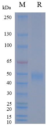
Purity
On SDS-PAGE under reducing (R) condition. The gel was stained overnight with Coomassie Blue. The purity of the protein is greater than 95%.
Affinity
Cynomolgus TNFR2 Protein; His Tag (Catalog # GM-88493RP) was immobilized at 2 μg/ml (100 μL/well). Increasing concentrations of Anti-H_TNFRSF1B(TNFR2) hIgG1 Antibody(UC2.3.8) (Catalog # GM-49245AB) were added.
Related products
For laboratory research use only. Direct human use, including taking orally and injection and clinical use are forbidden.
Cynomolgus TNFR2 Protein; His Tag
Cat. No.
GM-88493RP
Size
100 μg
1 mg
Quote
Description
Data Display
Related products
Description
Alternative Names TNFRSF1B, CD120b, TBPII, TNF-R-II, TNF-R75, TNFBR, TNFR1B, TNFR2
Source Cynomolgus TNFR2 Protein; His Tag (GM-88493RP) is expressed from human 293 cells (HEK-293). It contains AA Leu 23 - Asp 257 (Accession # XP_005544817.1).
This protein carries a His tag at the C-terminus.
Purity > 95% as determined by SDS-PAGE
Endotoxin < 1 EU/μg, determined by LAL gel clotting assay
Predicted Mol Mass 25.9 KDa
Formulation Supplied as a 0.2 μm filtered solution of PBS, pH7.2-7.4.
Description TNF receptors 1 and 2 (TNFR1 and TNFR2) mediate the effects of tumor necrosis factor (TNF). TNFR1, found on most nucleated cells, has a death domain and can trigger apoptosis or NF-κB/MAPK–mediated survival signals, activated by both soluble and membrane-bound TNF. TNFR2, mainly on immune and endothelial cells (lacks a death domain), promotes cell survival and regulatory T cell function via NF-κB and related pathways, often in response to membrane-bound TNF. The two receptors can form heterotrimers and modulate each other’s signaling, reflecting TNF biology’s complexity.
TNF-α signals primarily through its receptors TNFR1 (p55) and TNFR2 (p75), triggering multiple pathways. TNFR1 activation commonly leads to NF-κB and MAPK signaling, promoting transcription of inflammatory genes, survival factors, and sometimes apoptosis via caspase cascades. TNFR2 mainly modulates immune cell activation and can enhance TNF-α–driven responses. Cross-talk with other cytokines shapes the intensity and duration of inflammation. Therapeutically, TNF-α inhibitors are used to treat autoimmune diseases, but neutralization must balance infection risk and immune suppression.
Alternative Names TNFRSF1B, CD120b, TBPII, TNF-R-II, TNF-R75, TNFBR, TNFR1B, TNFR2
Source Cynomolgus TNFR2 Protein; His Tag (GM-88493RP) is expressed from human 293 cells (HEK-293). It contains AA Leu 23 - Asp 257 (Accession # XP_005544817.1).
This protein carries a His tag at the C-terminus.
Purity > 95% as determined by SDS-PAGE
Endotoxin < 1 EU/μg, determined by LAL gel clotting assay
Predicted Mol Mass 25.9 KDa
Formulation Supplied as a 0.2 μm filtered solution of PBS, pH7.2-7.4.
Description TNF receptors 1 and 2 (TNFR1 and TNFR2) mediate the effects of tumor necrosis factor (TNF). TNFR1, found on most nucleated cells, has a death domain and can trigger apoptosis or NF-κB/MAPK–mediated survival signals, activated by both soluble and membrane-bound TNF. TNFR2, mainly on immune and endothelial cells (lacks a death domain), promotes cell survival and regulatory T cell function via NF-κB and related pathways, often in response to membrane-bound TNF. The two receptors can form heterotrimers and modulate each other’s signaling, reflecting TNF biology’s complexity.
TNF-α signals primarily through its receptors TNFR1 (p55) and TNFR2 (p75), triggering multiple pathways. TNFR1 activation commonly leads to NF-κB and MAPK signaling, promoting transcription of inflammatory genes, survival factors, and sometimes apoptosis via caspase cascades. TNFR2 mainly modulates immune cell activation and can enhance TNF-α–driven responses. Cross-talk with other cytokines shapes the intensity and duration of inflammation. Therapeutically, TNF-α inhibitors are used to treat autoimmune diseases, but neutralization must balance infection risk and immune suppression.

Data Display
Purity
On SDS-PAGE under reducing (R) condition. The gel was stained overnight with Coomassie Blue. The purity of the protein is greater than 95%.
Affinity
Cynomolgus TNFR2 Protein; His Tag (Catalog # GM-88493RP) was immobilized at 2 μg/ml (100 μL/well). Increasing concentrations of Anti-H_TNFRSF1B(TNFR2) hIgG1 Antibody(UC2.3.8) (Catalog # GM-49245AB) were added.
Related products
Message consultation
reset
submit
Message
Message consultation
reset
submit