H_IL-1 Reporter 293 Cell Line
Cat. No.
GM-C43971
Size
1 vial
Quote
Description
Data Display
Specifications
Materials
Cell Culture
Related products
Description
Interleukin‑1 (IL‑1) is a key pro‑inflammatory cytokine with two forms, IL‑1α and IL‑1β, produced mainly by macrophages and also by endothelial and epithelial cells. Their activities are generally indistinguishable, and signaling occurs through IL‑1R1 (not IL‑1R2), which recruits IL‑1RAcP to form a high‑affinity receptor complex.

IL‑1α is rarely detected in circulation during disease, whereas IL‑1β is a major mediator of inflammation and host defense. Elevated IL‑1β is linked to atherosclerosis, type 2 diabetes, and autoimmune diseases such as rheumatoid arthritis, multiple sclerosis, and Crohn’s disease; blocking IL‑1 with a receptor antagonist can lessen autoinflammatory disease severity. IL‑1β synthesis, release, and activity are tightly controlled via innate sensing of PAMPs and DAMPs.

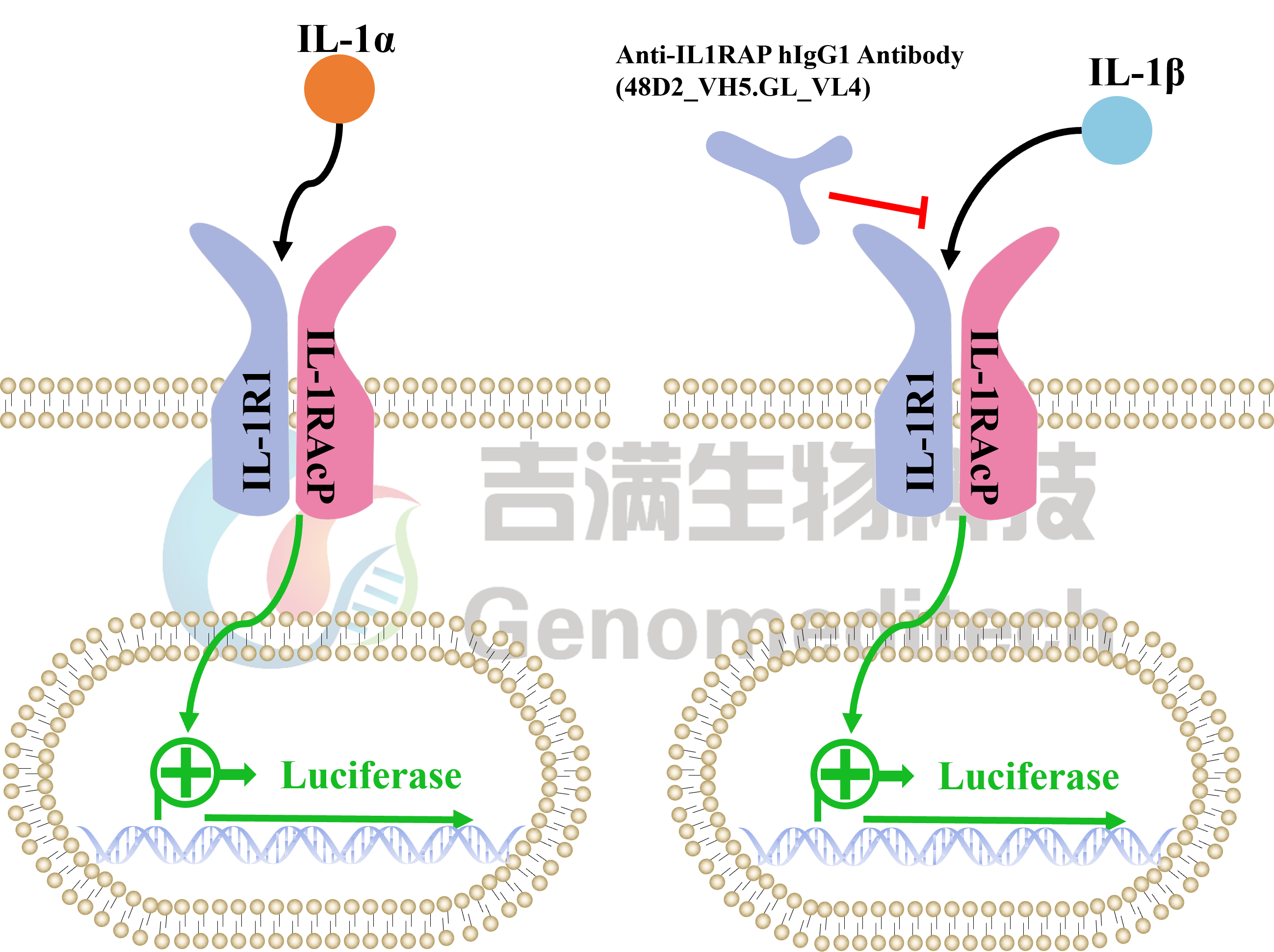
H_IL-1 Reporter 293 Cell Line is a clonal stable cell line with signal-dependent expression of a luciferase reporter gene constructed using lentiviral technology, and it endogenously expresses IL-1R1 and IL-1RAcP gene. When IL-1α or IL-1β binds to IL-1R1 and IL-1RAcP, it activates downstream signaling pathways, leading to the expression of luciferase. Blockade antibodies can inhibit this signal transmission. The luciferase activity measurement indicates the activation level of the signaling pathway and can thus be used to evaluate the in vitro effects of drugs related to IL-1.
Interleukin‑1 (IL‑1) is a key pro‑inflammatory cytokine with two forms, IL‑1α and IL‑1β, produced mainly by macrophages and also by endothelial and epithelial cells. Their activities are generally indistinguishable, and signaling occurs through IL‑1R1 (not IL‑1R2), which recruits IL‑1RAcP to form a high‑affinity receptor complex.

IL‑1α is rarely detected in circulation during disease, whereas IL‑1β is a major mediator of inflammation and host defense. Elevated IL‑1β is linked to atherosclerosis, type 2 diabetes, and autoimmune diseases such as rheumatoid arthritis, multiple sclerosis, and Crohn’s disease; blocking IL‑1 with a receptor antagonist can lessen autoinflammatory disease severity. IL‑1β synthesis, release, and activity are tightly controlled via innate sensing of PAMPs and DAMPs.

H_IL-1 Reporter 293 Cell Line is a clonal stable cell line with signal-dependent expression of a luciferase reporter gene constructed using lentiviral technology, and it endogenously expresses IL-1R1 and IL-1RAcP gene. When IL-1α or IL-1β binds to IL-1R1 and IL-1RAcP, it activates downstream signaling pathways, leading to the expression of luciferase. Blockade antibodies can inhibit this signal transmission. The luciferase activity measurement indicates the activation level of the signaling pathway and can thus be used to evaluate the in vitro effects of drugs related to IL-1.
Data Display
Signaling Pathway
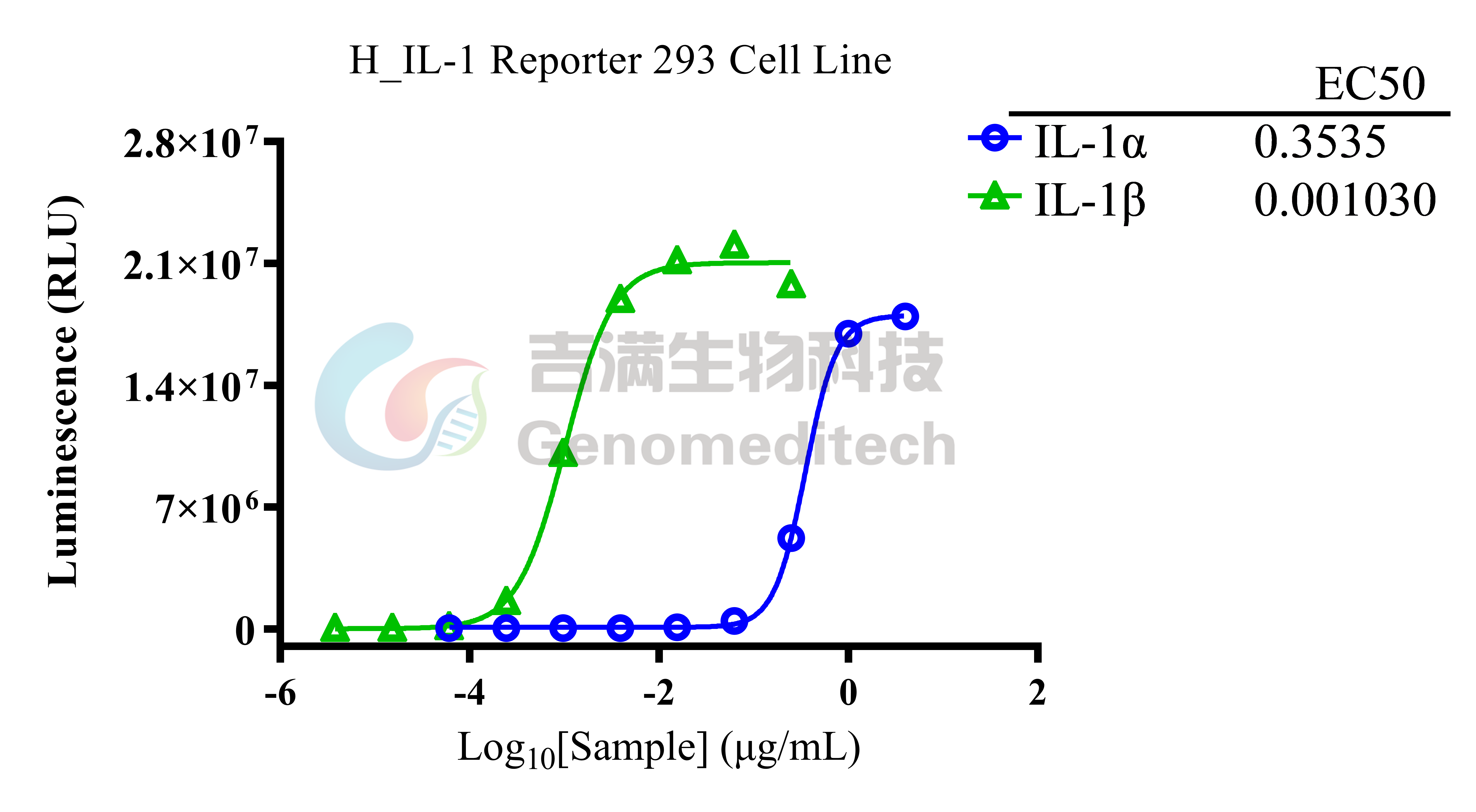
Response to human IL-1 alpha and IL-1 beta protein. The H_IL-1 Reporter 293 Cell Line (Cat. GM-C43971) at a concentration of 1.5E4 cells/well (96-well format) was stimulated with serial dilutions of Recombinant Human IL-1 alpha Protein (Cat. GM-88245RP), Human IL-1 beta Protein (Cat. GM-88248RP) in assay buffer (DMEM+1% FBS+1% P.S) for 6 hours. The firefly luciferase activity was measured using the Luciferase Reporter Assay Kit (Genomeditech). The maximum induction folds of IL-1α, IL-1β were approximately [687.4]and [584.6]. Data are shown by drug mass concentration.
Applications
Response to Anti-IL1RAP hIgG1 Antibody (48D2_VH5.GL_VL4). Serial dilutions of the Anti-IL1RAP hIgG1 Antibody (48D2_VH5.GL_VL4)(Cat. GM-88388AB) was incubated with 1.5E4 cells/well of the H_IL-1 Reporter 293 Cell Line (Cat. GM-C43971) in a 96-well plate for 1 hour in assay buffer (DMEM + 1% FBS + 1% P.S). Subsequently, the Human IL-1 beta Protein (Cat. GM-88248RP) at a concentration of 0.15 ng/well was added, and the coculture proceeded for an additional 6 hours. Firefly luciferase activity is then measured using the Luciferase Reporter Assay Kit (Genomeditech). The results indicated maximum blocking folds of approximately [101.8]. Data are shown by drug mass concentration.
Expression
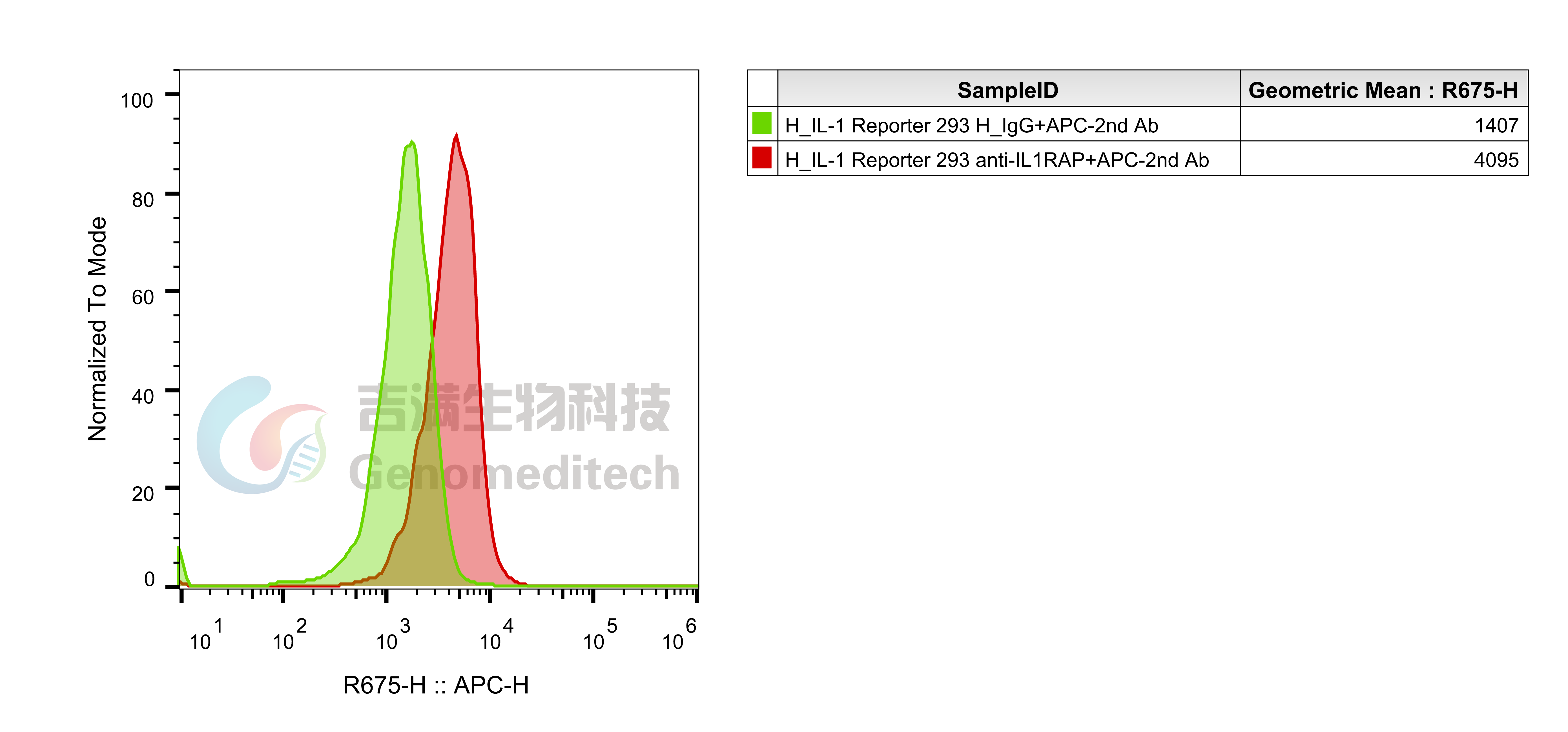
H_IL-1 Reporter 293 Cell Line (Cat. GM-C43971) was determined by flow cytometry using Anti-IL1RAP hIgG1 Antibody (48D2_VH5.GL_VL4)(Cat. GM-88388AB).
Specifications
Cat. No GM-C43971
Product H_IL-1 Reporter 293 Cell Line
Product Format 1 vial of frozen cells
Quantity 5E6 Cells per vial,1 mL
Storage Conditions Liquid nitrogen immediately upon receipt
Recovery Medium DMEM+10% FBS+1% P.S
Growth medium DMEM+10% FBS+1% P.S+4 μg/mL Blasticidin
Note None
Freezing Medium 90% FBS+10% DMSO
Growth properties Adherent
Growth Conditions 37°C, 5% CO₂
Safety considerations Biosafety Level 2
Mycoplasma Testing The cell line has been screened to confirm the absence of Mycoplasma species.
Cat. No GM-C43971
Product H_IL-1 Reporter 293 Cell Line
Product Format 1 vial of frozen cells
Quantity 5E6 Cells per vial,1 mL
Storage Conditions Liquid nitrogen immediately upon receipt
Recovery Medium DMEM+10% FBS+1% P.S
Growth medium DMEM+10% FBS+1% P.S+4 μg/mL Blasticidin
Note None
Freezing Medium 90% FBS+10% DMSO
Growth properties Adherent
Growth Conditions 37°C, 5% CO₂
Safety considerations Biosafety Level 2
Mycoplasma Testing The cell line has been screened to confirm the absence of Mycoplasma species.
Materials
Reagent Ordering Information
DMEM Gibco/C11995500BT
Fetal Bovine Serum ExCell/FSP500
Pen/Strep Thermo/15140-122
Blasticidin Genomeditech/GM-040404
Human IL-1 alpha Protein; His Tag Genomeditech/GM-88245RP
Anti-IL1RAP hIgG1 Antibody (48D2_VH5.GL_VL4) Genomeditech/GM-88388AB
Reagent Ordering Information
DMEM Gibco/C11995500BT
Fetal Bovine Serum ExCell/FSP500
Pen/Strep Thermo/15140-122
Blasticidin Genomeditech/GM-040404
Human IL-1 alpha Protein; His Tag Genomeditech/GM-88245RP
Anti-IL1RAP hIgG1 Antibody (48D2_VH5.GL_VL4) Genomeditech/GM-88388AB
Cell Culture

Cell Recovery

Recovery Medium: DMEM+10% FBS+1% P.S

To insure the highest level of viability, thaw the vial and initiate the culture as soon as possible upon receipt. If upon arrival, continued storage of the frozen culture is necessary, it should be stored in liquid nitrogen vapor phase and not at -70°C. Storage at -70°C will result in loss of viability.

a)       Thaw the vial by gentle agitation in a 37°C water bath. To reduce the possibility of contamination, keep the O-ring and cap out of the water. Thawing should be rapid (approximately 2 - 3 minutes).

b)       Remove the vial from the water bath as soon as the contents are thawed, and decontaminate by dipping in or spraying with 70% ethanol. All of the operations from this point on should be carried out under strict aseptic conditions.

c)       Transfer the vial contents to a centrifuge tube containing 5.0 mL complete culture medium. And spin at approximately 176 x g for 5 minutes. Discard supernatant.

d)       Resuspend cell pellet with the recommended complete medium. And dispense the suspension into an appropriate culture flask and initially place the flask in an upright position after thawing.

e)       Incubate the culture at 37°C in a suitable incubator. A 5% CO₂ in air atmosphere is recommended if using the medium described on this product sheet.

Cell Freezing

Freezing Medium: 90% FBS+10% DMSO

a)          Centrifuge at 176 x g for 3 minutes to collect cells.

b)         Resuspend the cells in pre-cooled freezing medium and adjust the cell density to 5E6 cells/mL.

c)          Aliquot 1 mL into each vial.

d)         Place the vial in a controlled-rate freezing container and store at -80°C for at least 1 day, then transfer to liquid nitrogen as soon as possible.

Cell passage

Growth medium: DMEM+10% FBS+1% P.S+4 μg/mL Blasticidin

For the first 1 to 2 passages post-resuscitation, use the recovery medium. Once the cells have stabilized, switch to a growth medium.

a)          Remove and discard culture medium.

b)         Briefly rinse the cell layer with PBS to remove all traces of serum that contains trypsin inhibitor.

c)          Add 1.0 mL of 0.25% (w/v) Trypsin-EDTA solution to dish and observe cells under an inverted microscope until cell layer is dispersed (usually within 30 to 60 seconds at 37°C).

d)         Note: To avoid clumping do not agitate the cells by hitting or shaking the flask while waiting for the cells to detach. Cells that are difficult to detach may be placed at 37°C to facilitate dispersal.

e)          Add 2.0 mL of growth medium to mix well and aspirate cells by gently pipetting.

f)          After centrifugation, resuspend the pellet and add appropriate aliquots of the cell suspension to new culture vessels.

g)         Incubate cultures at 37°C.

Subcultivation Ratio: A subcultivation ratio of 1:3 - 1:4 is recommended

Medium Renewal: Every 3 days

Notes

a)          After initial thawing, a higher proportion of dead cells is normal. The cell culture generally improves noticeably after approximately one week of adaptation. Once the culture stabilizes, the percentage of dead cells decreases with subsequent passages, and the cell proliferation rate becomes more consistent.

b)         It is important to maintain the cell density below 80%, as exceeding this threshold can lead to decreased cell viability and metabolic activity due to overcrowding.

c)          FBS requires heat inactivation at 56°C for 30 minutes, which can inactivate complement and some viruses, but does not significantly affect the activity of most growth factors and cytokines.

Cell Recovery

Recovery Medium: DMEM+10% FBS+1% P.S

To insure the highest level of viability, thaw the vial and initiate the culture as soon as possible upon receipt. If upon arrival, continued storage of the frozen culture is necessary, it should be stored in liquid nitrogen vapor phase and not at -70°C. Storage at -70°C will result in loss of viability.

a)       Thaw the vial by gentle agitation in a 37°C water bath. To reduce the possibility of contamination, keep the O-ring and cap out of the water. Thawing should be rapid (approximately 2 - 3 minutes).

b)       Remove the vial from the water bath as soon as the contents are thawed, and decontaminate by dipping in or spraying with 70% ethanol. All of the operations from this point on should be carried out under strict aseptic conditions.

c)       Transfer the vial contents to a centrifuge tube containing 5.0 mL complete culture medium. And spin at approximately 176 x g for 5 minutes. Discard supernatant.

d)       Resuspend cell pellet with the recommended complete medium. And dispense the suspension into an appropriate culture flask and initially place the flask in an upright position after thawing.

e)       Incubate the culture at 37°C in a suitable incubator. A 5% CO₂ in air atmosphere is recommended if using the medium described on this product sheet.

Cell Freezing

Freezing Medium: 90% FBS+10% DMSO

a)          Centrifuge at 176 x g for 3 minutes to collect cells.

b)         Resuspend the cells in pre-cooled freezing medium and adjust the cell density to 5E6 cells/mL.

c)          Aliquot 1 mL into each vial.

d)         Place the vial in a controlled-rate freezing container and store at -80°C for at least 1 day, then transfer to liquid nitrogen as soon as possible.

Cell passage

Growth medium: DMEM+10% FBS+1% P.S+4 μg/mL Blasticidin

For the first 1 to 2 passages post-resuscitation, use the recovery medium. Once the cells have stabilized, switch to a growth medium.

a)          Remove and discard culture medium.

b)         Briefly rinse the cell layer with PBS to remove all traces of serum that contains trypsin inhibitor.

c)          Add 1.0 mL of 0.25% (w/v) Trypsin-EDTA solution to dish and observe cells under an inverted microscope until cell layer is dispersed (usually within 30 to 60 seconds at 37°C).

d)         Note: To avoid clumping do not agitate the cells by hitting or shaking the flask while waiting for the cells to detach. Cells that are difficult to detach may be placed at 37°C to facilitate dispersal.

e)          Add 2.0 mL of growth medium to mix well and aspirate cells by gently pipetting.

f)          After centrifugation, resuspend the pellet and add appropriate aliquots of the cell suspension to new culture vessels.

g)         Incubate cultures at 37°C.

Subcultivation Ratio: A subcultivation ratio of 1:3 - 1:4 is recommended

Medium Renewal: Every 3 days

Notes

a)          After initial thawing, a higher proportion of dead cells is normal. The cell culture generally improves noticeably after approximately one week of adaptation. Once the culture stabilizes, the percentage of dead cells decreases with subsequent passages, and the cell proliferation rate becomes more consistent.

b)         It is important to maintain the cell density below 80%, as exceeding this threshold can lead to decreased cell viability and metabolic activity due to overcrowding.

c)          FBS requires heat inactivation at 56°C for 30 minutes, which can inactivate complement and some viruses, but does not significantly affect the activity of most growth factors and cytokines.

For laboratory research use only. Direct human use, including taking orally and injection and clinical use are forbidden.
H_IL-1 Reporter 293 Cell Line
Cat. No.
GM-C43971
Size
1 vial
Quote
Description
Data Display
Specifications
Materials
Cell Culture
Related products
Description
Interleukin‑1 (IL‑1) is a key pro‑inflammatory cytokine with two forms, IL‑1α and IL‑1β, produced mainly by macrophages and also by endothelial and epithelial cells. Their activities are generally indistinguishable, and signaling occurs through IL‑1R1 (not IL‑1R2), which recruits IL‑1RAcP to form a high‑affinity receptor complex.

IL‑1α is rarely detected in circulation during disease, whereas IL‑1β is a major mediator of inflammation and host defense. Elevated IL‑1β is linked to atherosclerosis, type 2 diabetes, and autoimmune diseases such as rheumatoid arthritis, multiple sclerosis, and Crohn’s disease; blocking IL‑1 with a receptor antagonist can lessen autoinflammatory disease severity. IL‑1β synthesis, release, and activity are tightly controlled via innate sensing of PAMPs and DAMPs.

H_IL-1 Reporter 293 Cell Line is a clonal stable cell line with signal-dependent expression of a luciferase reporter gene constructed using lentiviral technology, and it endogenously expresses IL-1R1 and IL-1RAcP gene. When IL-1α or IL-1β binds to IL-1R1 and IL-1RAcP, it activates downstream signaling pathways, leading to the expression of luciferase. Blockade antibodies can inhibit this signal transmission. The luciferase activity measurement indicates the activation level of the signaling pathway and can thus be used to evaluate the in vitro effects of drugs related to IL-1.

Interleukin‑1 (IL‑1) is a key pro‑inflammatory cytokine with two forms, IL‑1α and IL‑1β, produced mainly by macrophages and also by endothelial and epithelial cells. Their activities are generally indistinguishable, and signaling occurs through IL‑1R1 (not IL‑1R2), which recruits IL‑1RAcP to form a high‑affinity receptor complex.

IL‑1α is rarely detected in circulation during disease, whereas IL‑1β is a major mediator of inflammation and host defense. Elevated IL‑1β is linked to atherosclerosis, type 2 diabetes, and autoimmune diseases such as rheumatoid arthritis, multiple sclerosis, and Crohn’s disease; blocking IL‑1 with a receptor antagonist can lessen autoinflammatory disease severity. IL‑1β synthesis, release, and activity are tightly controlled via innate sensing of PAMPs and DAMPs.

H_IL-1 Reporter 293 Cell Line is a clonal stable cell line with signal-dependent expression of a luciferase reporter gene constructed using lentiviral technology, and it endogenously expresses IL-1R1 and IL-1RAcP gene. When IL-1α or IL-1β binds to IL-1R1 and IL-1RAcP, it activates downstream signaling pathways, leading to the expression of luciferase. Blockade antibodies can inhibit this signal transmission. The luciferase activity measurement indicates the activation level of the signaling pathway and can thus be used to evaluate the in vitro effects of drugs related to IL-1.

Data Display
Signaling Pathway
Response to human IL-1 alpha and IL-1 beta protein. The H_IL-1 Reporter 293 Cell Line (Cat. GM-C43971) at a concentration of 1.5E4 cells/well (96-well format) was stimulated with serial dilutions of Recombinant Human IL-1 alpha Protein (Cat. GM-88245RP), Human IL-1 beta Protein (Cat. GM-88248RP) in assay buffer (DMEM+1% FBS+1% P.S) for 6 hours. The firefly luciferase activity was measured using the Luciferase Reporter Assay Kit (Genomeditech). The maximum induction folds of IL-1α, IL-1β were approximately [687.4]and [584.6]. Data are shown by drug mass concentration.
Applications
Response to Anti-IL1RAP hIgG1 Antibody (48D2_VH5.GL_VL4). Serial dilutions of the Anti-IL1RAP hIgG1 Antibody (48D2_VH5.GL_VL4)(Cat. GM-88388AB) was incubated with 1.5E4 cells/well of the H_IL-1 Reporter 293 Cell Line (Cat. GM-C43971) in a 96-well plate for 1 hour in assay buffer (DMEM + 1% FBS + 1% P.S). Subsequently, the Human IL-1 beta Protein (Cat. GM-88248RP) at a concentration of 0.15 ng/well was added, and the coculture proceeded for an additional 6 hours. Firefly luciferase activity is then measured using the Luciferase Reporter Assay Kit (Genomeditech). The results indicated maximum blocking folds of approximately [101.8]. Data are shown by drug mass concentration.
Expression
H_IL-1 Reporter 293 Cell Line (Cat. GM-C43971) was determined by flow cytometry using Anti-IL1RAP hIgG1 Antibody (48D2_VH5.GL_VL4)(Cat. GM-88388AB).
Specifications
Cat. No GM-C43971
Product H_IL-1 Reporter 293 Cell Line
Product Format 1 vial of frozen cells
Quantity 5E6 Cells per vial,1 mL
Storage Conditions Liquid nitrogen immediately upon receipt
Recovery Medium DMEM+10% FBS+1% P.S
Growth medium DMEM+10% FBS+1% P.S+4 μg/mL Blasticidin
Note None
Freezing Medium 90% FBS+10% DMSO
Growth properties Adherent
Growth Conditions 37°C, 5% CO₂
Safety considerations Biosafety Level 2
Mycoplasma Testing The cell line has been screened to confirm the absence of Mycoplasma species.
Cat. No GM-C43971
Product H_IL-1 Reporter 293 Cell Line
Product Format 1 vial of frozen cells
Quantity 5E6 Cells per vial,1 mL
Storage Conditions Liquid nitrogen immediately upon receipt
Recovery Medium DMEM+10% FBS+1% P.S
Growth medium DMEM+10% FBS+1% P.S+4 μg/mL Blasticidin
Note None
Freezing Medium 90% FBS+10% DMSO
Growth properties Adherent
Growth Conditions 37°C, 5% CO₂
Safety considerations Biosafety Level 2
Mycoplasma Testing The cell line has been screened to confirm the absence of Mycoplasma species.
Materials
Reagent Ordering Information
DMEM Gibco/C11995500BT
Fetal Bovine Serum ExCell/FSP500
Pen/Strep Thermo/15140-122
Blasticidin Genomeditech/GM-040404
Human IL-1 alpha Protein; His Tag Genomeditech/GM-88245RP
Anti-IL1RAP hIgG1 Antibody (48D2_VH5.GL_VL4) Genomeditech/GM-88388AB
Reagent Ordering Information
DMEM Gibco/C11995500BT
Fetal Bovine Serum ExCell/FSP500
Pen/Strep Thermo/15140-122
Blasticidin Genomeditech/GM-040404
Human IL-1 alpha Protein; His Tag Genomeditech/GM-88245RP
Anti-IL1RAP hIgG1 Antibody (48D2_VH5.GL_VL4) Genomeditech/GM-88388AB
Cell Culture

Cell Recovery

Recovery Medium: DMEM+10% FBS+1% P.S

To insure the highest level of viability, thaw the vial and initiate the culture as soon as possible upon receipt. If upon arrival, continued storage of the frozen culture is necessary, it should be stored in liquid nitrogen vapor phase and not at -70°C. Storage at -70°C will result in loss of viability.

a)       Thaw the vial by gentle agitation in a 37°C water bath. To reduce the possibility of contamination, keep the O-ring and cap out of the water. Thawing should be rapid (approximately 2 - 3 minutes).

b)       Remove the vial from the water bath as soon as the contents are thawed, and decontaminate by dipping in or spraying with 70% ethanol. All of the operations from this point on should be carried out under strict aseptic conditions.

c)       Transfer the vial contents to a centrifuge tube containing 5.0 mL complete culture medium. And spin at approximately 176 x g for 5 minutes. Discard supernatant.

d)       Resuspend cell pellet with the recommended complete medium. And dispense the suspension into an appropriate culture flask and initially place the flask in an upright position after thawing.

e)       Incubate the culture at 37°C in a suitable incubator. A 5% CO₂ in air atmosphere is recommended if using the medium described on this product sheet.

Cell Freezing

Freezing Medium: 90% FBS+10% DMSO

a)          Centrifuge at 176 x g for 3 minutes to collect cells.

b)         Resuspend the cells in pre-cooled freezing medium and adjust the cell density to 5E6 cells/mL.

c)          Aliquot 1 mL into each vial.

d)         Place the vial in a controlled-rate freezing container and store at -80°C for at least 1 day, then transfer to liquid nitrogen as soon as possible.

Cell passage

Growth medium: DMEM+10% FBS+1% P.S+4 μg/mL Blasticidin

For the first 1 to 2 passages post-resuscitation, use the recovery medium. Once the cells have stabilized, switch to a growth medium.

a)          Remove and discard culture medium.

b)         Briefly rinse the cell layer with PBS to remove all traces of serum that contains trypsin inhibitor.

c)          Add 1.0 mL of 0.25% (w/v) Trypsin-EDTA solution to dish and observe cells under an inverted microscope until cell layer is dispersed (usually within 30 to 60 seconds at 37°C).

d)         Note: To avoid clumping do not agitate the cells by hitting or shaking the flask while waiting for the cells to detach. Cells that are difficult to detach may be placed at 37°C to facilitate dispersal.

e)          Add 2.0 mL of growth medium to mix well and aspirate cells by gently pipetting.

f)          After centrifugation, resuspend the pellet and add appropriate aliquots of the cell suspension to new culture vessels.

g)         Incubate cultures at 37°C.

Subcultivation Ratio: A subcultivation ratio of 1:3 - 1:4 is recommended

Medium Renewal: Every 3 days

Notes

a)          After initial thawing, a higher proportion of dead cells is normal. The cell culture generally improves noticeably after approximately one week of adaptation. Once the culture stabilizes, the percentage of dead cells decreases with subsequent passages, and the cell proliferation rate becomes more consistent.

b)         It is important to maintain the cell density below 80%, as exceeding this threshold can lead to decreased cell viability and metabolic activity due to overcrowding.

c)          FBS requires heat inactivation at 56°C for 30 minutes, which can inactivate complement and some viruses, but does not significantly affect the activity of most growth factors and cytokines.

Cell Recovery

Recovery Medium: DMEM+10% FBS+1% P.S

To insure the highest level of viability, thaw the vial and initiate the culture as soon as possible upon receipt. If upon arrival, continued storage of the frozen culture is necessary, it should be stored in liquid nitrogen vapor phase and not at -70°C. Storage at -70°C will result in loss of viability.

a)       Thaw the vial by gentle agitation in a 37°C water bath. To reduce the possibility of contamination, keep the O-ring and cap out of the water. Thawing should be rapid (approximately 2 - 3 minutes).

b)       Remove the vial from the water bath as soon as the contents are thawed, and decontaminate by dipping in or spraying with 70% ethanol. All of the operations from this point on should be carried out under strict aseptic conditions.

c)       Transfer the vial contents to a centrifuge tube containing 5.0 mL complete culture medium. And spin at approximately 176 x g for 5 minutes. Discard supernatant.

d)       Resuspend cell pellet with the recommended complete medium. And dispense the suspension into an appropriate culture flask and initially place the flask in an upright position after thawing.

e)       Incubate the culture at 37°C in a suitable incubator. A 5% CO₂ in air atmosphere is recommended if using the medium described on this product sheet.

Cell Freezing

Freezing Medium: 90% FBS+10% DMSO

a)          Centrifuge at 176 x g for 3 minutes to collect cells.

b)         Resuspend the cells in pre-cooled freezing medium and adjust the cell density to 5E6 cells/mL.

c)          Aliquot 1 mL into each vial.

d)         Place the vial in a controlled-rate freezing container and store at -80°C for at least 1 day, then transfer to liquid nitrogen as soon as possible.

Cell passage

Growth medium: DMEM+10% FBS+1% P.S+4 μg/mL Blasticidin

For the first 1 to 2 passages post-resuscitation, use the recovery medium. Once the cells have stabilized, switch to a growth medium.

a)          Remove and discard culture medium.

b)         Briefly rinse the cell layer with PBS to remove all traces of serum that contains trypsin inhibitor.

c)          Add 1.0 mL of 0.25% (w/v) Trypsin-EDTA solution to dish and observe cells under an inverted microscope until cell layer is dispersed (usually within 30 to 60 seconds at 37°C).

d)         Note: To avoid clumping do not agitate the cells by hitting or shaking the flask while waiting for the cells to detach. Cells that are difficult to detach may be placed at 37°C to facilitate dispersal.

e)          Add 2.0 mL of growth medium to mix well and aspirate cells by gently pipetting.

f)          After centrifugation, resuspend the pellet and add appropriate aliquots of the cell suspension to new culture vessels.

g)         Incubate cultures at 37°C.

Subcultivation Ratio: A subcultivation ratio of 1:3 - 1:4 is recommended

Medium Renewal: Every 3 days

Notes

a)          After initial thawing, a higher proportion of dead cells is normal. The cell culture generally improves noticeably after approximately one week of adaptation. Once the culture stabilizes, the percentage of dead cells decreases with subsequent passages, and the cell proliferation rate becomes more consistent.

b)         It is important to maintain the cell density below 80%, as exceeding this threshold can lead to decreased cell viability and metabolic activity due to overcrowding.

c)          FBS requires heat inactivation at 56°C for 30 minutes, which can inactivate complement and some viruses, but does not significantly affect the activity of most growth factors and cytokines.

Message consultation
reset
submit
Message
Message consultation
reset
submit