Where Discovery Begins
H_AR Reporter 293 Cell Line
Cat. No.
GM-C45341
Size
2 vial
Quote
Description
Data Display
Specifications
Materials
Cell Culture
Related products
Description
Androgen receptor (AR), also known as NR3C4 and encoded by the AR gene, is a ligand-activated transcription factor belonging to the steroid receptor subfamily of the nuclear receptor superfamily, predominantly expressed in androgen-responsive tissues such as the prostate, testis, and skeletal muscle. As the specific receptor for testosterone and dihydrotestosterone (DHT), AR plays a pivotal role in male sexual differentiation, prostate development, and bone metabolism, making it a critical therapeutic target for prostate cancer, androgenetic alopecia, and muscle-wasting disorders.

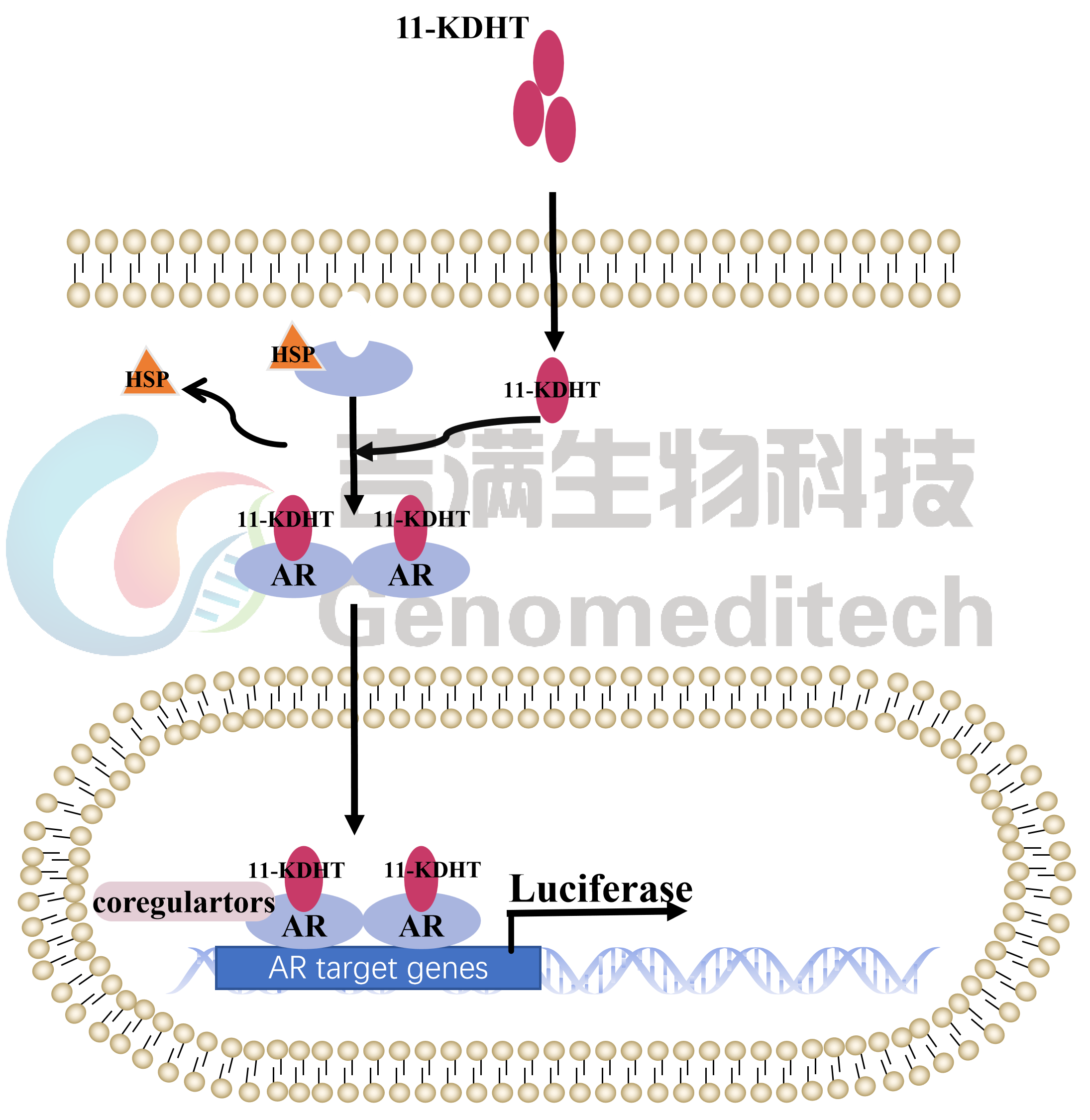
Upon binding of DHT or testosterone, AR undergoes conformational change, dissociates from heat shock proteins, dimerizes, and translocates into the nucleus, where it binds to androgen response elements (AREs) to activate target gene transcription.driver of prostate cancer progression.

H_AR Reporter Cell Line is a clonal stable cell line constructed using lentiviral technology, constitutive expression of the AR gene, along with signal-dependent expression of a luciferase reporter gene under the control of androgen response elements (AREs). When DHT or other AR agonists bind to AR, it undergoes nuclear translocation and binds to AREs, activating the expression of luciferase. The luciferase activity measurement indicates the activation level of the AR signaling pathway and can thus be used to evaluate the in vitro effects of drugs related to AR.
Androgen receptor (AR), also known as NR3C4 and encoded by the AR gene, is a ligand-activated transcription factor belonging to the steroid receptor subfamily of the nuclear receptor superfamily, predominantly expressed in androgen-responsive tissues such as the prostate, testis, and skeletal muscle. As the specific receptor for testosterone and dihydrotestosterone (DHT), AR plays a pivotal role in male sexual differentiation, prostate development, and bone metabolism, making it a critical therapeutic target for prostate cancer, androgenetic alopecia, and muscle-wasting disorders.

Upon binding of DHT or testosterone, AR undergoes conformational change, dissociates from heat shock proteins, dimerizes, and translocates into the nucleus, where it binds to androgen response elements (AREs) to activate target gene transcription.driver of prostate cancer progression.

H_AR Reporter Cell Line is a clonal stable cell line constructed using lentiviral technology, constitutive expression of the AR gene, along with signal-dependent expression of a luciferase reporter gene under the control of androgen response elements (AREs). When DHT or other AR agonists bind to AR, it undergoes nuclear translocation and binds to AREs, activating the expression of luciferase. The luciferase activity measurement indicates the activation level of the AR signaling pathway and can thus be used to evaluate the in vitro effects of drugs related to AR.
Data Display
Signaling Pathway
Response to 11-Ketodihydrotestosterone and Metribolone. The H_AR Reporter 293 Cell Line (Cat. GM-C45341) at a density of 1.5E4 cells/well (96-well format) were stimulated with serial dilutions of 11-Ketodihydrotestosterone(MCE/HY-135794) and Metribolone (GLPBIO/GC19800) in assay buffer (DMEM + 1% FBS + 1% P.S) for 6 hours. The firefly luciferase activity was measured using the Luciferase Reporter Assay Kit (Genomeditech). The maximum induction fold was approximately [33.8] and [28.0]. Data are shown by drug molar concentration.
Applications
Inhibition of 11-Ketodihydrotestosterone induced reporter activity by Bicalutamide. Serial dilutions of the Bicalutamide(MCE/HY-14249) was incubated with 1.5E4 cells/well of the H_AR Reporter 293 Cell Line (Cat. GM-C45341) in a 96-well plate for 1 hour in assay buffer (DMEM +1% FBS+1% P.S). Subsequently, the 11-Ketodihydrotestosterone (MCE/HY-135794) was added to each well at a final concentration of 3 nM,, and the coculture proceeded for an additional 6 hours. Firefly luciferase activity is then measured using the Luciferase Reporter Assay Kit (Genomeditech)(left Y-axis, relative luminescence units), with inhibition percentages shown on the right Y-axis.
Inhibition of 11-Ketodihydrotestosterone induced reporter activity by Enzalutamide. Serial dilutions of the Enzalutamide(MCE/HY-70002) was incubated with 1.5E4 cells/well of the H_AR Reporter 293 Cell Line (Cat. GM-C45341) in a 96-well plate for 1 hour in assay buffer (DMEM +1% FBS+1% P.S). Subsequently, the 11-Ketodihydrotestosterone (MCE/HY-135794) was added to each well at a final concentration of 3 nM,, and the coculture proceeded for an additional 6 hours. Firefly luciferase activity is then measured using the Luciferase Reporter Assay Kit (Genomeditech)(left Y-axis, relative luminescence units), with inhibition percentages shown on the right Y-axis.
Expression
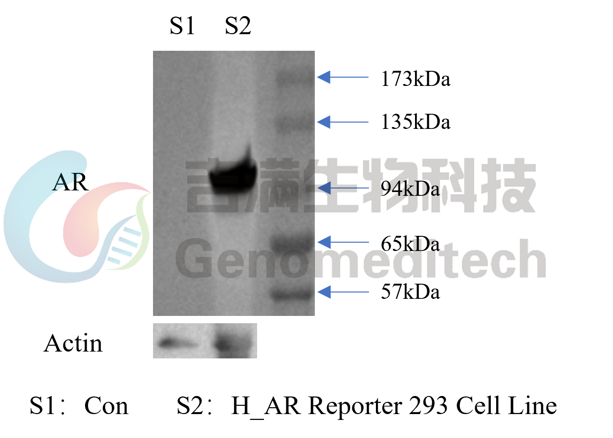
The protein expression levels of AR in the H_AR Reporter 293 Cell Line(Cat. GM-C45341 were determined by Western blotting (WB).
Specifications
Cat. No GM-C45341
Product H_AR Reporter 293 Cell Line
Product Format 1 vial of frozen cells
Quantity 5E6 Cells per vial,1 mL
Storage Conditions Liquid nitrogen immediately upon receipt
Recovery Medium DMEM+10% FBS+1% P.S
Growth medium DMEM+10% FBS+1% P.S+4 μg/mL Blasticidin+0.75 μg/mL Puromycin
Note None
Freezing Medium 90% FBS+10% DMSO
Growth properties Adherent
Growth Conditions 37°C, 5% CO₂
Safety considerations Biosafety Level 2
Mycoplasma Testing The cell line has been screened to confirm the absence of Mycoplasma species.
Cat. No GM-C45341
Product H_AR Reporter 293 Cell Line
Product Format 1 vial of frozen cells
Quantity 5E6 Cells per vial,1 mL
Storage Conditions Liquid nitrogen immediately upon receipt
Recovery Medium DMEM+10% FBS+1% P.S
Growth medium DMEM+10% FBS+1% P.S+4 μg/mL Blasticidin+0.75 μg/mL Puromycin
Note None
Freezing Medium 90% FBS+10% DMSO
Growth properties Adherent
Growth Conditions 37°C, 5% CO₂
Safety considerations Biosafety Level 2
Mycoplasma Testing The cell line has been screened to confirm the absence of Mycoplasma species.
Materials
Reagent Ordering Information
DMEM Gibco/C11995500BT
Fetal Bovine Serum ExCell/FSP500
Pen/Strep Thermo/15140-122
Blasticidin Genomeditech/GM-040404
Puromycin Genomeditech/GM-040401
Androgen Receptor(D6F11) Rabbit Monoclonal Antibody CST/#5153
11-Ketodihydrotestosterone MCE/HY-135794
Metribolone GLPBIO/GC19800
Bicalutamide MCE/HY-14249
Enzalutamide MCE/HY-70002
Reagent Ordering Information
DMEM Gibco/C11995500BT
Fetal Bovine Serum ExCell/FSP500
Pen/Strep Thermo/15140-122
Blasticidin Genomeditech/GM-040404
Puromycin Genomeditech/GM-040401
Androgen Receptor(D6F11) Rabbit Monoclonal Antibody CST/#5153
11-Ketodihydrotestosterone MCE/HY-135794
Metribolone GLPBIO/GC19800
Bicalutamide MCE/HY-14249
Enzalutamide MCE/HY-70002
Cell Culture

Cell Recovery

Recovery Medium: DMEM+10% FBS+1% P.S

To insure the highest level of viability, thaw the vial and initiate the culture as soon as possible upon receipt. If upon arrival, continued storage of the frozen culture is necessary, it should be stored in liquid nitrogen vapor phase and not at -70°C. Storage at -70°C will result in loss of viability.

a)       Thaw the vial by gentle agitation in a 37°C water bath. To reduce the possibility of contamination, keep the O-ring and cap out of the water. Thawing should be rapid (approximately 2 - 3 minutes).

b)       Remove the vial from the water bath as soon as the contents are thawed, and decontaminate by dipping in or spraying with 70% ethanol. All of the operations from this point on should be carried out under strict aseptic conditions.

c)       Transfer the vial contents to a centrifuge tube containing 5.0 mL complete culture medium and spin at approximately 176 x g for 5 minutes. Discard supernatant.

d)       Resuspend cell pellet with the recommended recovery medium. And dispense into appropriate culture dishes.

e)       Incubate the culture at 37°C in a suitable incubator. A 5% CO₂ in air atmosphere is recommended if using the medium described on this product sheet.

Cell Freezing

Freezing Medium: 90% FBS+10% DMSO

a)          Centrifuge at 176 x g for 3 minutes to collect cells.

b)         Resuspend the cells in pre-cooled freezing medium and adjust the cell density to 5E6 cells/mL.

c)          Aliquot 1 mL into each vial.

d)         Place the vial in a controlled-rate freezing container and store at -80°C for at least 1 day, then transfer to liquid nitrogen as soon as possible.

Cell passage

Growth medium: DMEM+10% FBS+1% P.S+4 μg/mL Blasticidin+0.75 μg/mL Puromycin

For the first 1 to 2 passages post-resuscitation, use the recovery medium. Once the cells have stabilized, switch to a growth medium.

a)          Subculturing is necessary when the cell density reaches 80%. It is recommended to perform subculturing at a ratio of 1:3 to 1:4 every 2-3 days. Ensure that the density does not exceed 80%, as overcrowding can lead to reduced viability due to compression.

b)         Remove and discard culture medium.

c)          Briefly rinse the cell layer with PBS to remove all traces of serum that contains trypsin inhibitor.

d)         Add 1.0 mL of 0.25% (w/v) Trypsin-EDTA solution to dish and observe cells under an inverted microscope until cell layer is dispersed (usually within 30 to 60 seconds at 37°C).

e)          Note: To avoid clumping do not agitate the cells by hitting or shaking the flask while waiting for the cells to detach. Cells that are difficult to detach may be placed at 37°C to facilitate dispersal.

f)          Add 2.0 mL of growth medium to mix well and aspirate cells by gently pipetting.

g)         After centrifugation, resuspend the pellet and add appropriate aliquots of the cell suspension to new culture vessels.

h)         Incubate cultures at 37°C.

Subcultivation Ratio: A subcultivation ratio of 1:3 - 1:4 is recommended

Medium Renewal: Every 2 to 3 days

Notes

a)          Upon initial thawing, a higher number of dead cells is observed, which is a normal phenomenon. Significant improvement is seen after adaptation. Once the cells reach a stable state, the number of dead cells decreases after subculturing and the cell growth rate becomes stable.

b)         Ensure that the cell density does not exceed 80%, as overcrowding may lead to reduced viability due to compression.

Cell Recovery

Recovery Medium: DMEM+10% FBS+1% P.S

To insure the highest level of viability, thaw the vial and initiate the culture as soon as possible upon receipt. If upon arrival, continued storage of the frozen culture is necessary, it should be stored in liquid nitrogen vapor phase and not at -70°C. Storage at -70°C will result in loss of viability.

a)       Thaw the vial by gentle agitation in a 37°C water bath. To reduce the possibility of contamination, keep the O-ring and cap out of the water. Thawing should be rapid (approximately 2 - 3 minutes).

b)       Remove the vial from the water bath as soon as the contents are thawed, and decontaminate by dipping in or spraying with 70% ethanol. All of the operations from this point on should be carried out under strict aseptic conditions.

c)       Transfer the vial contents to a centrifuge tube containing 5.0 mL complete culture medium and spin at approximately 176 x g for 5 minutes. Discard supernatant.

d)       Resuspend cell pellet with the recommended recovery medium. And dispense into appropriate culture dishes.

e)       Incubate the culture at 37°C in a suitable incubator. A 5% CO₂ in air atmosphere is recommended if using the medium described on this product sheet.

Cell Freezing

Freezing Medium: 90% FBS+10% DMSO

a)          Centrifuge at 176 x g for 3 minutes to collect cells.

b)         Resuspend the cells in pre-cooled freezing medium and adjust the cell density to 5E6 cells/mL.

c)          Aliquot 1 mL into each vial.

d)         Place the vial in a controlled-rate freezing container and store at -80°C for at least 1 day, then transfer to liquid nitrogen as soon as possible.

Cell passage

Growth medium: DMEM+10% FBS+1% P.S+4 μg/mL Blasticidin+0.75 μg/mL Puromycin

For the first 1 to 2 passages post-resuscitation, use the recovery medium. Once the cells have stabilized, switch to a growth medium.

a)          Subculturing is necessary when the cell density reaches 80%. It is recommended to perform subculturing at a ratio of 1:3 to 1:4 every 2-3 days. Ensure that the density does not exceed 80%, as overcrowding can lead to reduced viability due to compression.

b)         Remove and discard culture medium.

c)          Briefly rinse the cell layer with PBS to remove all traces of serum that contains trypsin inhibitor.

d)         Add 1.0 mL of 0.25% (w/v) Trypsin-EDTA solution to dish and observe cells under an inverted microscope until cell layer is dispersed (usually within 30 to 60 seconds at 37°C).

e)          Note: To avoid clumping do not agitate the cells by hitting or shaking the flask while waiting for the cells to detach. Cells that are difficult to detach may be placed at 37°C to facilitate dispersal.

f)          Add 2.0 mL of growth medium to mix well and aspirate cells by gently pipetting.

g)         After centrifugation, resuspend the pellet and add appropriate aliquots of the cell suspension to new culture vessels.

h)         Incubate cultures at 37°C.

Subcultivation Ratio: A subcultivation ratio of 1:3 - 1:4 is recommended

Medium Renewal: Every 2 to 3 days

Notes

a)          Upon initial thawing, a higher number of dead cells is observed, which is a normal phenomenon. Significant improvement is seen after adaptation. Once the cells reach a stable state, the number of dead cells decreases after subculturing and the cell growth rate becomes stable.

b)         Ensure that the cell density does not exceed 80%, as overcrowding may lead to reduced viability due to compression.

Related products
FOLH1(PSMA)
Cynomolgus_FOLH1(PSMA) CHO-K1 Cell Line H_FOLH1(PSMA) CHO-K1 Cell Line H_FOLH1(PSMA) HEK-293 Cell Line
H_FOLH1(PSMA) PC-3 Cell Line H_FOLH1(PSMA) RM-1 Cell Line
Anti-FOLH1(PSMA) hIgG1 Antibody Anti-FOLH1(PSMA) hIgG1 Reference Antibody (Rosobio)
Cynomolgus FOLH1(PSMA) Protein; His Tag Human FOLH1(PSMA) Protein; His Tag  
ADC Related Product
Anti-DXD Mouse IgG1 Antibody (23E21C5) Anti-DXD Mouse IgG1 Antibody (4A5A12) Anti-Dxd Mouse IgG2a Antibody (17D6A4)
Anti-Eribulin Mouse IgG2a Antibody (10F8G4) Anti-MMAE Mouse IgG1 Antibody (11C10E3) Anti-MMAE Mouse IgG2a Antibody (17A1K11)
Anti-MMAE Mouse IgG2a Antibody (8F6A3) Anti-SN38 Mouse IgG1 Antibody(59H11C7) Mouse anti Human IgG1-DXD(Dar8)
Mouse anti Human IgG1-MMAE(Dar4) Human IgG1 Isotype-DXD (Dar8) Human IgG1 Isotype-Eribulin (Dar4)
Human IgG1 Isotype-MMAE (Dar4)
Recombinant DT3C Protein    
AR(NR3C4)
H_AR HEK-293 Cell Line    
KLK2 KLK3
Membrane bound H_KLK2(AA19-261) 22RV1 Cell Line Membrane bound H_KLK2(AA19-261) CHO-K1 Cell Line Membrane bound H_KLK2(AA19-261) HEK-293 Cell Line
Membrane bound H_KLK2(AA19-261) LnCap Cell Line Membrane bound H_KLK2(AA25-261) CHO-K1 Cell Line Membrane bound H_KLK2(AA25-261) CT26 Cell Line
Membrane bound H_KLK2(AA25-261) HEK-293 Cell Line Membrane bound H_KLK2(AA25-261) MC38 Cell Line (Low Expression)
Anti-KLK2 hIgG1 Antibody(Hu11B6)
Biotinylated Human KLK2 Protein; His-Avi Tag    
ACP3
Flag-H_ACP3 HCT116 Cell Line Flag-H_ACP3 HT-1080 Cell Line H_ACP3 CHO-K1 Cell Line
H_ACP3 HCT116 Cell Line H_ACP3 HEK-293 Cell Line H_ACP3 HT-1080 Cell Line
H_ACP3 PC-3 Cell Line    
For laboratory research use only. Direct human use, including taking orally and injection and clinical use are forbidden.
H_AR Reporter 293 Cell Line
Cat. No.
GM-C45341
Size
2 vial
Quote
Description
Data Display
Specifications
Materials
Cell Culture
Related products
Description
Androgen receptor (AR), also known as NR3C4 and encoded by the AR gene, is a ligand-activated transcription factor belonging to the steroid receptor subfamily of the nuclear receptor superfamily, predominantly expressed in androgen-responsive tissues such as the prostate, testis, and skeletal muscle. As the specific receptor for testosterone and dihydrotestosterone (DHT), AR plays a pivotal role in male sexual differentiation, prostate development, and bone metabolism, making it a critical therapeutic target for prostate cancer, androgenetic alopecia, and muscle-wasting disorders.

Upon binding of DHT or testosterone, AR undergoes conformational change, dissociates from heat shock proteins, dimerizes, and translocates into the nucleus, where it binds to androgen response elements (AREs) to activate target gene transcription.driver of prostate cancer progression.

H_AR Reporter Cell Line is a clonal stable cell line constructed using lentiviral technology, constitutive expression of the AR gene, along with signal-dependent expression of a luciferase reporter gene under the control of androgen response elements (AREs). When DHT or other AR agonists bind to AR, it undergoes nuclear translocation and binds to AREs, activating the expression of luciferase. The luciferase activity measurement indicates the activation level of the AR signaling pathway and can thus be used to evaluate the in vitro effects of drugs related to AR.

Androgen receptor (AR), also known as NR3C4 and encoded by the AR gene, is a ligand-activated transcription factor belonging to the steroid receptor subfamily of the nuclear receptor superfamily, predominantly expressed in androgen-responsive tissues such as the prostate, testis, and skeletal muscle. As the specific receptor for testosterone and dihydrotestosterone (DHT), AR plays a pivotal role in male sexual differentiation, prostate development, and bone metabolism, making it a critical therapeutic target for prostate cancer, androgenetic alopecia, and muscle-wasting disorders.

Upon binding of DHT or testosterone, AR undergoes conformational change, dissociates from heat shock proteins, dimerizes, and translocates into the nucleus, where it binds to androgen response elements (AREs) to activate target gene transcription.driver of prostate cancer progression.

H_AR Reporter Cell Line is a clonal stable cell line constructed using lentiviral technology, constitutive expression of the AR gene, along with signal-dependent expression of a luciferase reporter gene under the control of androgen response elements (AREs). When DHT or other AR agonists bind to AR, it undergoes nuclear translocation and binds to AREs, activating the expression of luciferase. The luciferase activity measurement indicates the activation level of the AR signaling pathway and can thus be used to evaluate the in vitro effects of drugs related to AR.

Data Display
Signaling Pathway
Response to 11-Ketodihydrotestosterone and Metribolone. The H_AR Reporter 293 Cell Line (Cat. GM-C45341) at a density of 1.5E4 cells/well (96-well format) were stimulated with serial dilutions of 11-Ketodihydrotestosterone(MCE/HY-135794) and Metribolone (GLPBIO/GC19800) in assay buffer (DMEM + 1% FBS + 1% P.S) for 6 hours. The firefly luciferase activity was measured using the Luciferase Reporter Assay Kit (Genomeditech). The maximum induction fold was approximately [33.8] and [28.0]. Data are shown by drug molar concentration.
Applications
Inhibition of 11-Ketodihydrotestosterone induced reporter activity by Bicalutamide. Serial dilutions of the Bicalutamide(MCE/HY-14249) was incubated with 1.5E4 cells/well of the H_AR Reporter 293 Cell Line (Cat. GM-C45341) in a 96-well plate for 1 hour in assay buffer (DMEM +1% FBS+1% P.S). Subsequently, the 11-Ketodihydrotestosterone (MCE/HY-135794) was added to each well at a final concentration of 3 nM,, and the coculture proceeded for an additional 6 hours. Firefly luciferase activity is then measured using the Luciferase Reporter Assay Kit (Genomeditech)(left Y-axis, relative luminescence units), with inhibition percentages shown on the right Y-axis.
Inhibition of 11-Ketodihydrotestosterone induced reporter activity by Enzalutamide. Serial dilutions of the Enzalutamide(MCE/HY-70002) was incubated with 1.5E4 cells/well of the H_AR Reporter 293 Cell Line (Cat. GM-C45341) in a 96-well plate for 1 hour in assay buffer (DMEM +1% FBS+1% P.S). Subsequently, the 11-Ketodihydrotestosterone (MCE/HY-135794) was added to each well at a final concentration of 3 nM,, and the coculture proceeded for an additional 6 hours. Firefly luciferase activity is then measured using the Luciferase Reporter Assay Kit (Genomeditech)(left Y-axis, relative luminescence units), with inhibition percentages shown on the right Y-axis.
Expression
The protein expression levels of AR in the H_AR Reporter 293 Cell Line(Cat. GM-C45341 were determined by Western blotting (WB).
Specifications
Cat. No GM-C45341
Product H_AR Reporter 293 Cell Line
Product Format 1 vial of frozen cells
Quantity 5E6 Cells per vial,1 mL
Storage Conditions Liquid nitrogen immediately upon receipt
Recovery Medium DMEM+10% FBS+1% P.S
Growth medium DMEM+10% FBS+1% P.S+4 μg/mL Blasticidin+0.75 μg/mL Puromycin
Note None
Freezing Medium 90% FBS+10% DMSO
Growth properties Adherent
Growth Conditions 37°C, 5% CO₂
Safety considerations Biosafety Level 2
Mycoplasma Testing The cell line has been screened to confirm the absence of Mycoplasma species.
Cat. No GM-C45341
Product H_AR Reporter 293 Cell Line
Product Format 1 vial of frozen cells
Quantity 5E6 Cells per vial,1 mL
Storage Conditions Liquid nitrogen immediately upon receipt
Recovery Medium DMEM+10% FBS+1% P.S
Growth medium DMEM+10% FBS+1% P.S+4 μg/mL Blasticidin+0.75 μg/mL Puromycin
Note None
Freezing Medium 90% FBS+10% DMSO
Growth properties Adherent
Growth Conditions 37°C, 5% CO₂
Safety considerations Biosafety Level 2
Mycoplasma Testing The cell line has been screened to confirm the absence of Mycoplasma species.
Materials
Reagent Ordering Information
DMEM Gibco/C11995500BT
Fetal Bovine Serum ExCell/FSP500
Pen/Strep Thermo/15140-122
Blasticidin Genomeditech/GM-040404
Puromycin Genomeditech/GM-040401
Androgen Receptor(D6F11) Rabbit Monoclonal Antibody CST/#5153
11-Ketodihydrotestosterone MCE/HY-135794
Metribolone GLPBIO/GC19800
Bicalutamide MCE/HY-14249
Enzalutamide MCE/HY-70002
Reagent Ordering Information
DMEM Gibco/C11995500BT
Fetal Bovine Serum ExCell/FSP500
Pen/Strep Thermo/15140-122
Blasticidin Genomeditech/GM-040404
Puromycin Genomeditech/GM-040401
Androgen Receptor(D6F11) Rabbit Monoclonal Antibody CST/#5153
11-Ketodihydrotestosterone MCE/HY-135794
Metribolone GLPBIO/GC19800
Bicalutamide MCE/HY-14249
Enzalutamide MCE/HY-70002
Cell Culture

Cell Recovery

Recovery Medium: DMEM+10% FBS+1% P.S

To insure the highest level of viability, thaw the vial and initiate the culture as soon as possible upon receipt. If upon arrival, continued storage of the frozen culture is necessary, it should be stored in liquid nitrogen vapor phase and not at -70°C. Storage at -70°C will result in loss of viability.

a)       Thaw the vial by gentle agitation in a 37°C water bath. To reduce the possibility of contamination, keep the O-ring and cap out of the water. Thawing should be rapid (approximately 2 - 3 minutes).

b)       Remove the vial from the water bath as soon as the contents are thawed, and decontaminate by dipping in or spraying with 70% ethanol. All of the operations from this point on should be carried out under strict aseptic conditions.

c)       Transfer the vial contents to a centrifuge tube containing 5.0 mL complete culture medium and spin at approximately 176 x g for 5 minutes. Discard supernatant.

d)       Resuspend cell pellet with the recommended recovery medium. And dispense into appropriate culture dishes.

e)       Incubate the culture at 37°C in a suitable incubator. A 5% CO₂ in air atmosphere is recommended if using the medium described on this product sheet.

Cell Freezing

Freezing Medium: 90% FBS+10% DMSO

a)          Centrifuge at 176 x g for 3 minutes to collect cells.

b)         Resuspend the cells in pre-cooled freezing medium and adjust the cell density to 5E6 cells/mL.

c)          Aliquot 1 mL into each vial.

d)         Place the vial in a controlled-rate freezing container and store at -80°C for at least 1 day, then transfer to liquid nitrogen as soon as possible.

Cell passage

Growth medium: DMEM+10% FBS+1% P.S+4 μg/mL Blasticidin+0.75 μg/mL Puromycin

For the first 1 to 2 passages post-resuscitation, use the recovery medium. Once the cells have stabilized, switch to a growth medium.

a)          Subculturing is necessary when the cell density reaches 80%. It is recommended to perform subculturing at a ratio of 1:3 to 1:4 every 2-3 days. Ensure that the density does not exceed 80%, as overcrowding can lead to reduced viability due to compression.

b)         Remove and discard culture medium.

c)          Briefly rinse the cell layer with PBS to remove all traces of serum that contains trypsin inhibitor.

d)         Add 1.0 mL of 0.25% (w/v) Trypsin-EDTA solution to dish and observe cells under an inverted microscope until cell layer is dispersed (usually within 30 to 60 seconds at 37°C).

e)          Note: To avoid clumping do not agitate the cells by hitting or shaking the flask while waiting for the cells to detach. Cells that are difficult to detach may be placed at 37°C to facilitate dispersal.

f)          Add 2.0 mL of growth medium to mix well and aspirate cells by gently pipetting.

g)         After centrifugation, resuspend the pellet and add appropriate aliquots of the cell suspension to new culture vessels.

h)         Incubate cultures at 37°C.

Subcultivation Ratio: A subcultivation ratio of 1:3 - 1:4 is recommended

Medium Renewal: Every 2 to 3 days

Notes

a)          Upon initial thawing, a higher number of dead cells is observed, which is a normal phenomenon. Significant improvement is seen after adaptation. Once the cells reach a stable state, the number of dead cells decreases after subculturing and the cell growth rate becomes stable.

b)         Ensure that the cell density does not exceed 80%, as overcrowding may lead to reduced viability due to compression.


Cell Recovery

Recovery Medium: DMEM+10% FBS+1% P.S

To insure the highest level of viability, thaw the vial and initiate the culture as soon as possible upon receipt. If upon arrival, continued storage of the frozen culture is necessary, it should be stored in liquid nitrogen vapor phase and not at -70°C. Storage at -70°C will result in loss of viability.

a)       Thaw the vial by gentle agitation in a 37°C water bath. To reduce the possibility of contamination, keep the O-ring and cap out of the water. Thawing should be rapid (approximately 2 - 3 minutes).

b)       Remove the vial from the water bath as soon as the contents are thawed, and decontaminate by dipping in or spraying with 70% ethanol. All of the operations from this point on should be carried out under strict aseptic conditions.

c)       Transfer the vial contents to a centrifuge tube containing 5.0 mL complete culture medium and spin at approximately 176 x g for 5 minutes. Discard supernatant.

d)       Resuspend cell pellet with the recommended recovery medium. And dispense into appropriate culture dishes.

e)       Incubate the culture at 37°C in a suitable incubator. A 5% CO₂ in air atmosphere is recommended if using the medium described on this product sheet.


Cell Freezing

Freezing Medium: 90% FBS+10% DMSO

a)          Centrifuge at 176 x g for 3 minutes to collect cells.

b)         Resuspend the cells in pre-cooled freezing medium and adjust the cell density to 5E6 cells/mL.

c)          Aliquot 1 mL into each vial.

d)         Place the vial in a controlled-rate freezing container and store at -80°C for at least 1 day, then transfer to liquid nitrogen as soon as possible.

Cell passage

Growth medium: DMEM+10% FBS+1% P.S+4 μg/mL Blasticidin+0.75 μg/mL Puromycin

For the first 1 to 2 passages post-resuscitation, use the recovery medium. Once the cells have stabilized, switch to a growth medium.

a)          Subculturing is necessary when the cell density reaches 80%. It is recommended to perform subculturing at a ratio of 1:3 to 1:4 every 2-3 days. Ensure that the density does not exceed 80%, as overcrowding can lead to reduced viability due to compression.

b)         Remove and discard culture medium.

c)          Briefly rinse the cell layer with PBS to remove all traces of serum that contains trypsin inhibitor.

d)         Add 1.0 mL of 0.25% (w/v) Trypsin-EDTA solution to dish and observe cells under an inverted microscope until cell layer is dispersed (usually within 30 to 60 seconds at 37°C).

e)          Note: To avoid clumping do not agitate the cells by hitting or shaking the flask while waiting for the cells to detach. Cells that are difficult to detach may be placed at 37°C to facilitate dispersal.

f)          Add 2.0 mL of growth medium to mix well and aspirate cells by gently pipetting.

g)         After centrifugation, resuspend the pellet and add appropriate aliquots of the cell suspension to new culture vessels.

h)         Incubate cultures at 37°C.

Subcultivation Ratio: A subcultivation ratio of 1:3 - 1:4 is recommended

Medium Renewal: Every 2 to 3 days

Notes

a)          Upon initial thawing, a higher number of dead cells is observed, which is a normal phenomenon. Significant improvement is seen after adaptation. Once the cells reach a stable state, the number of dead cells decreases after subculturing and the cell growth rate becomes stable.


b)         Ensure that the cell density does not exceed 80%, as overcrowding may lead to reduced viability due to compression.

Related products
FOLH1(PSMA)
Cynomolgus_FOLH1(PSMA) CHO-K1 Cell Line H_FOLH1(PSMA) CHO-K1 Cell Line H_FOLH1(PSMA) HEK-293 Cell Line
H_FOLH1(PSMA) PC-3 Cell Line H_FOLH1(PSMA) RM-1 Cell Line
Anti-FOLH1(PSMA) hIgG1 Antibody Anti-FOLH1(PSMA) hIgG1 Reference Antibody (Rosobio)
Cynomolgus FOLH1(PSMA) Protein; His Tag Human FOLH1(PSMA) Protein; His Tag  
ADC Related Product
Anti-DXD Mouse IgG1 Antibody (23E21C5) Anti-DXD Mouse IgG1 Antibody (4A5A12) Anti-Dxd Mouse IgG2a Antibody (17D6A4)
Anti-Eribulin Mouse IgG2a Antibody (10F8G4) Anti-MMAE Mouse IgG1 Antibody (11C10E3) Anti-MMAE Mouse IgG2a Antibody (17A1K11)
Anti-MMAE Mouse IgG2a Antibody (8F6A3) Anti-SN38 Mouse IgG1 Antibody(59H11C7) Mouse anti Human IgG1-DXD(Dar8)
Mouse anti Human IgG1-MMAE(Dar4) Human IgG1 Isotype-DXD (Dar8) Human IgG1 Isotype-Eribulin (Dar4)
Human IgG1 Isotype-MMAE (Dar4)
Recombinant DT3C Protein    
AR(NR3C4)
H_AR HEK-293 Cell Line    
KLK2 KLK3
Membrane bound H_KLK2(AA19-261) 22RV1 Cell Line Membrane bound H_KLK2(AA19-261) CHO-K1 Cell Line Membrane bound H_KLK2(AA19-261) HEK-293 Cell Line
Membrane bound H_KLK2(AA19-261) LnCap Cell Line Membrane bound H_KLK2(AA25-261) CHO-K1 Cell Line Membrane bound H_KLK2(AA25-261) CT26 Cell Line
Membrane bound H_KLK2(AA25-261) HEK-293 Cell Line Membrane bound H_KLK2(AA25-261) MC38 Cell Line (Low Expression)
Anti-KLK2 hIgG1 Antibody(Hu11B6)
Biotinylated Human KLK2 Protein; His-Avi Tag    
ACP3
Flag-H_ACP3 HCT116 Cell Line Flag-H_ACP3 HT-1080 Cell Line H_ACP3 CHO-K1 Cell Line
H_ACP3 HCT116 Cell Line H_ACP3 HEK-293 Cell Line H_ACP3 HT-1080 Cell Line
H_ACP3 PC-3 Cell Line    
Contact
Us
Message Inquiry
reset
submit
Message
Message Inquiry
reset
submit