Where Discovery Begins
Cynomolgus_CD3D CD3E Reporter Jurkat(CD3DE KO) Cell
Cat. No.
GM-C45147
Size
2 vial
Quote
Description
Data Display
Specifications
Materials
Cell Culture
Related products
Description
CD3D and CD3E form the CD3δε heterodimer, a core subunit of the TCR‑CD3 complex essential for proper assembly and surface expression. Upon TCR engagement, their cytoplasmic ITAMs are phosphorylated, triggering downstream signaling that drives T‑cell activation, proliferation, and effector functions. CD3D is critical for thymocyte development, while CD3E regulates receptor internalization. As a major target in bispecific antibody development (over 400 active programs), most CD3‑targeting antibodies recognize CD3ε and mediate antitumor effects through T‑cell engagement. Therefore, a cell model expressing cynomolgus CD3 is valuable for preclinical cross‑reactivity and pharmacodynamic assessment.

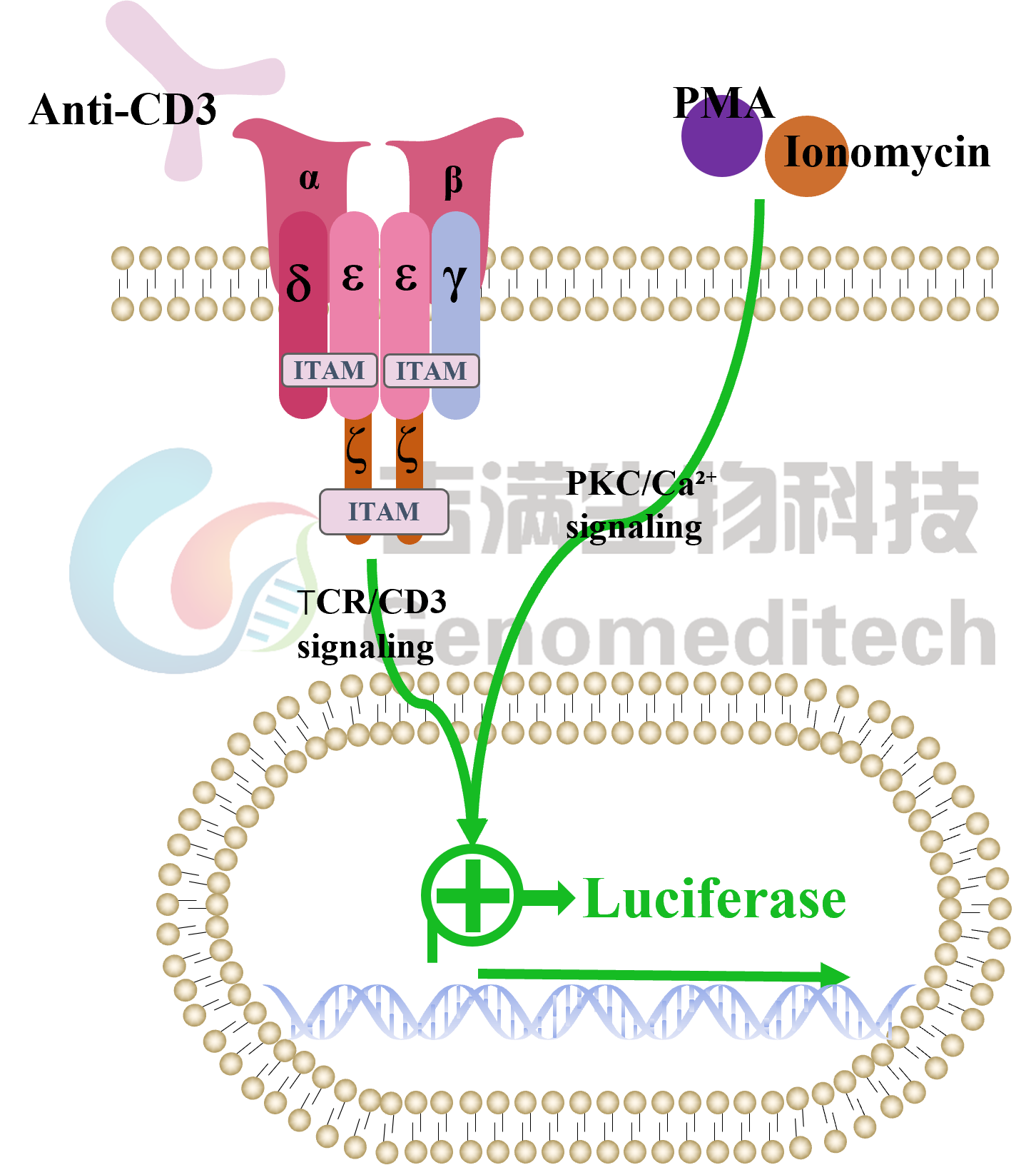
Cynomolgus_CD3D CD3E Reporter Jurkat(CD3DE KO) Cell Line is a clonal stable cell line that knockout CD3D/E and constitutive expression of Cynomolgus_CD3D/E gene, along with signal-dependent expression of a luciferase reporter gene. When drugs capable of binding and activating cynomolgus CD3 are added, the TCR‑CD3 complex becomes cross‑linked, triggering downstream signaling pathways that drive luciferase expression. The luciferase readout reflects the extent of pathway activation and can be used to evaluate the agonistic activity or functional binding of candidate drugs to cynomolgus CD3.
CD3D and CD3E form the CD3δε heterodimer, a core subunit of the TCR‑CD3 complex essential for proper assembly and surface expression. Upon TCR engagement, their cytoplasmic ITAMs are phosphorylated, triggering downstream signaling that drives T‑cell activation, proliferation, and effector functions. CD3D is critical for thymocyte development, while CD3E regulates receptor internalization. As a major target in bispecific antibody development (over 400 active programs), most CD3‑targeting antibodies recognize CD3ε and mediate antitumor effects through T‑cell engagement. Therefore, a cell model expressing cynomolgus CD3 is valuable for preclinical cross‑reactivity and pharmacodynamic assessment.

Cynomolgus_CD3D CD3E Reporter Jurkat(CD3DE KO) Cell Line is a clonal stable cell line that knockout CD3D/E and constitutive expression of Cynomolgus_CD3D/E gene, along with signal-dependent expression of a luciferase reporter gene. When drugs capable of binding and activating cynomolgus CD3 are added, the TCR‑CD3 complex becomes cross‑linked, triggering downstream signaling pathways that drive luciferase expression. The luciferase readout reflects the extent of pathway activation and can be used to evaluate the agonistic activity or functional binding of candidate drugs to cynomolgus CD3.
Data Display
Signaling Pathway
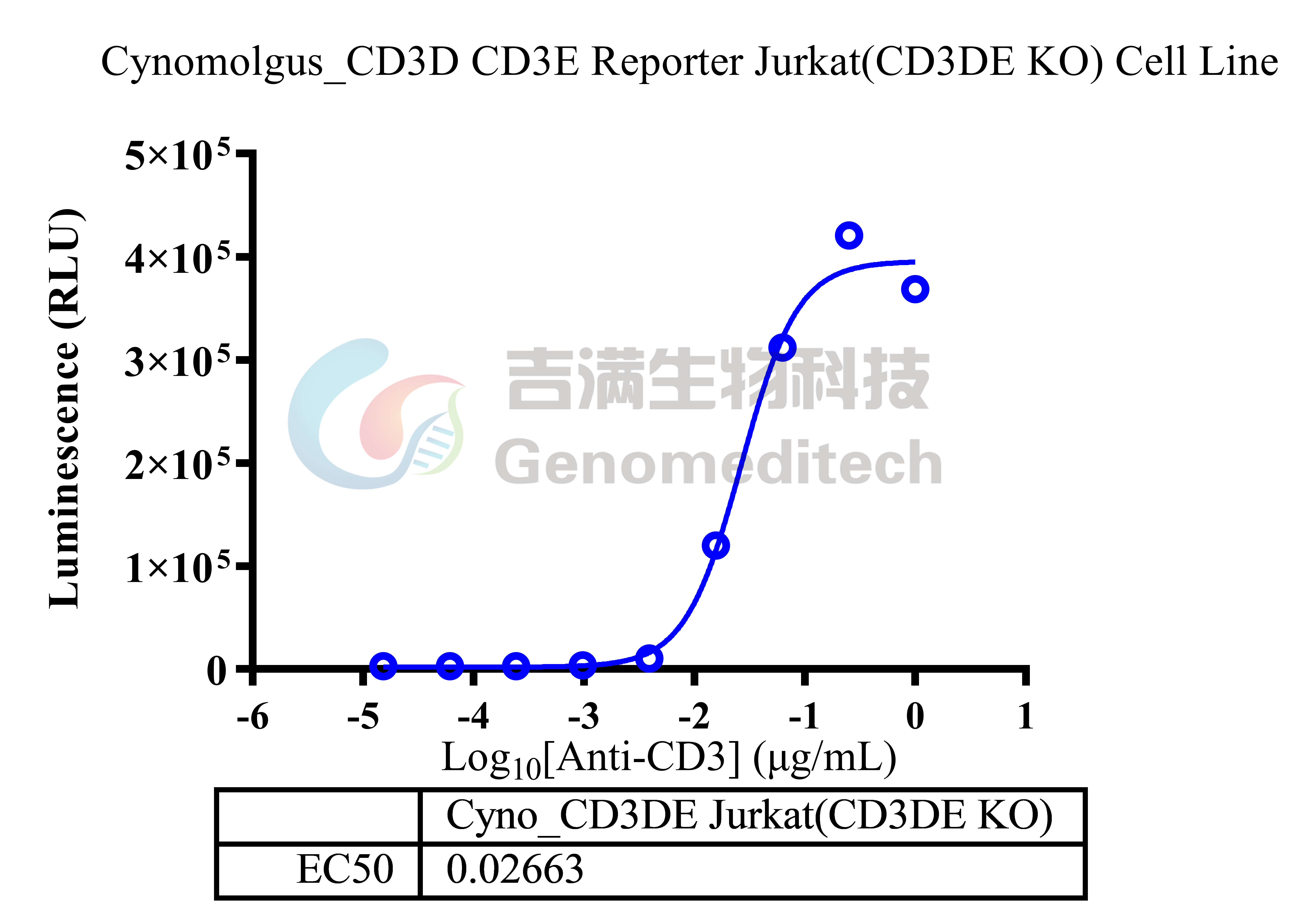
Response to Anti-CD3 hIgG1 Antibody(CH2527). The Cynomolgus_CD3D CD3E Reporter Jurkat(CD3DE KO) Cell Line (Cat. GM-C45147) at a concentration of 5E4 cells/well (96-well format) was stimulated with serial dilutions of Anti-CD3 hIgG1 Antibody(CH2527) (Cat. GM-33037AB) in assay buffer (RPMI 1640+1% FBS+1% P.S) for 6 hours. The firefly luciferase activity was measured using the Luciferase Reporter Assay Kit (Genomeditech). The maximum induction fold was approximately [126.1]. Data are shown by drug mass concentration.
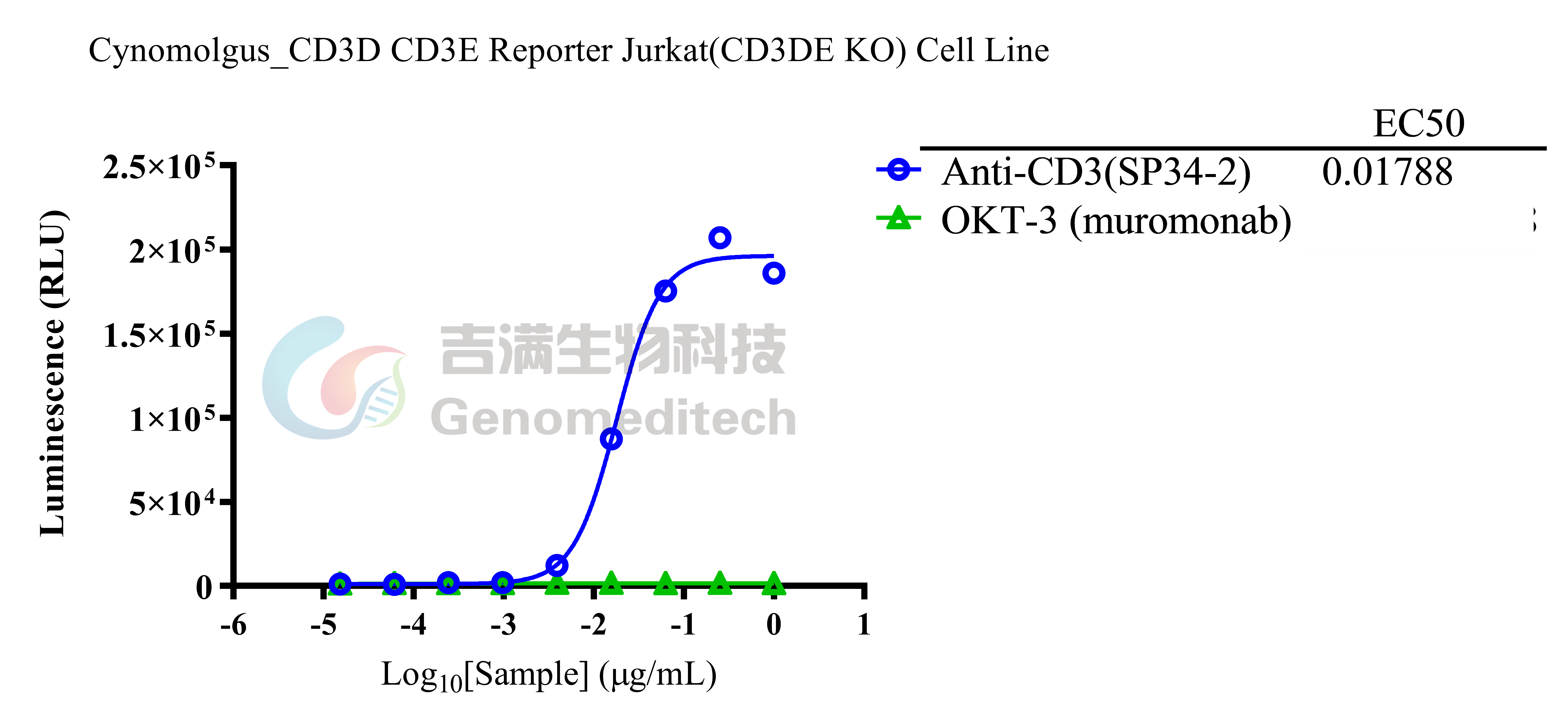
Response to Anti-CD3 mIgG1 Antibody(SP34-2). The Cynomolgus_CD3D CD3E Reporter Jurkat(CD3DE KO) Cell Line (Cat. GM-C45147) at a concentration of 5E4 cells/well (96-well format) was stimulated with serial dilutions of Anti-CD3 mIgG1 Antibody(SP34-2) (Cat. GM-87919AB) and Anti-CD3 epsilon Antibody (Cat. GM-51478AB) in assay buffer (RPMI 1640+1% FBS+1% P.S) for 6 hours. The firefly luciferase activity was measured using the Luciferase Reporter Assay Kit (Genomeditech). The firefly luciferase activity was measured using the Luciferase Reporter Assay Kit (Genomeditech). The maximum induction fold was approximately [136.6]. Data are shown by drug mass concentration.
Response to PMA/TPA. The Cynomolgus_CD3D CD3E Reporter Jurkat(CD3DE KO) Cell Line (Cat. GM-C45147) at a concentration of 5E4 cells/well (96-well format) was stimulated with serial dilutions of PMA/TPA (Beyotime/S1819) and 500nM Ionomycin (MCE/HY-13434) in assay buffer (RPMI 1640+1% FBS+1% P.S) for 6 hours. The firefly luciferase activity was measured using the Luciferase Reporter Assay Kit (Genomeditech). The maximum induction fold was approximately [9.7]. Data are shown by drug molar concentration.
Expression
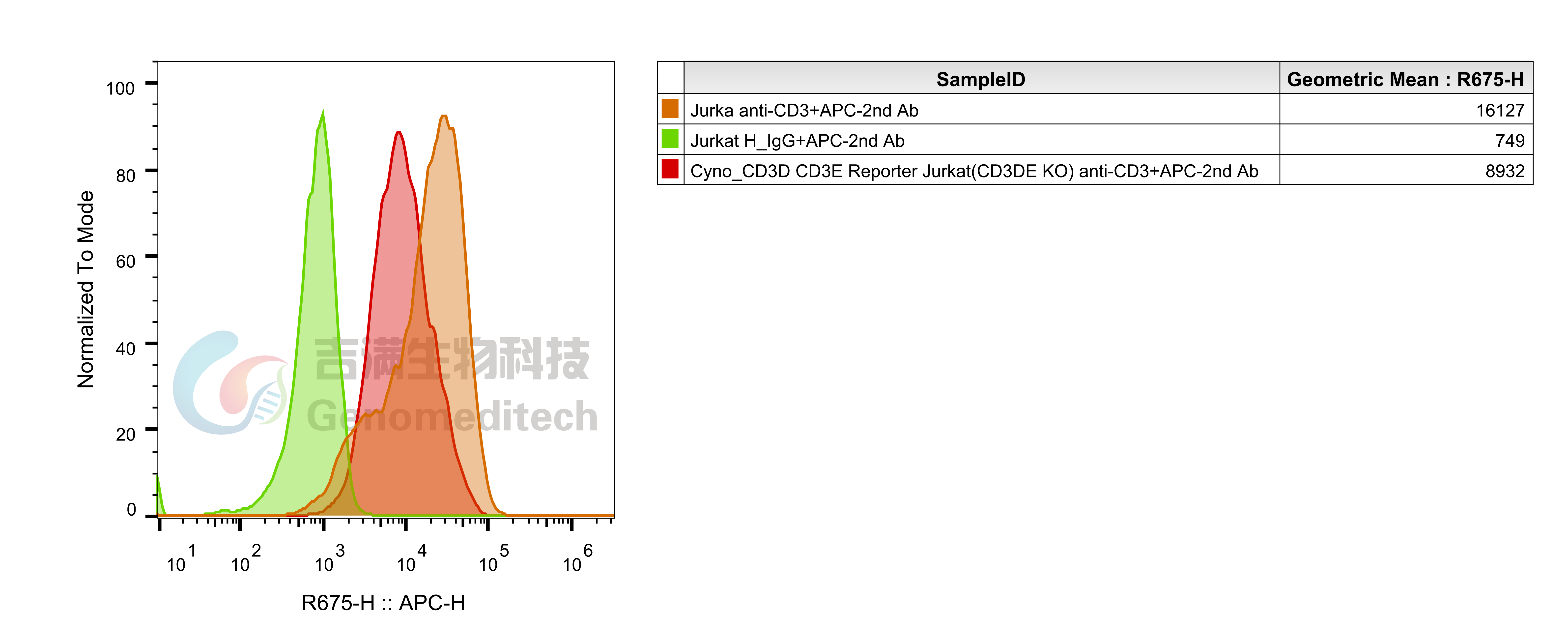
Cynomolgus_CD3D CD3E Reporter Jurkat(CD3DE KO) Cell Line (Cat. GM-C45147) was determined by flow cytometry using Anti-CD3 hIgG1 Antibody(CH2527)(Cat. GM-33037AB).
Genotype Verification
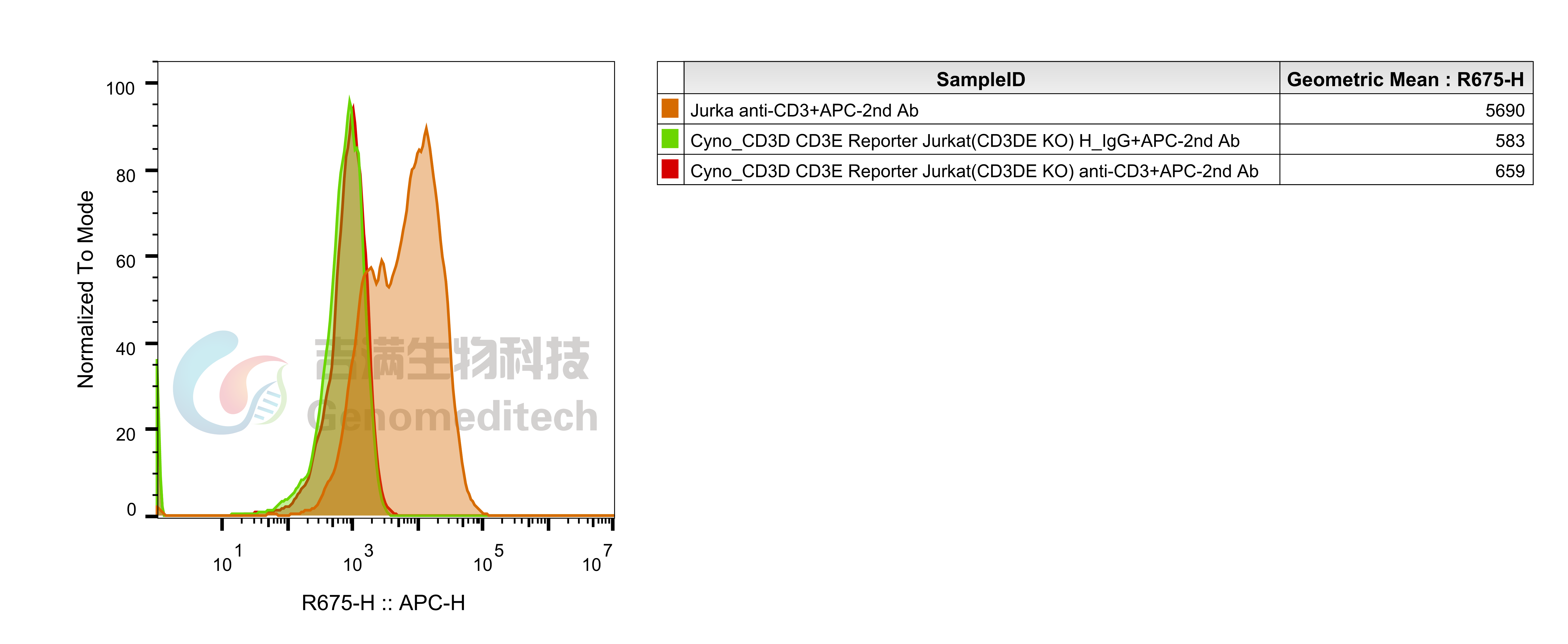
Cynomolgus_CD3D CD3E Reporter Jurkat(CD3DE KO) Cell Line (Cat. GM-C45147) was determined by flow cytometry using Anti-CD3 epsilon hIgG1 Antibody (Cat. GM-51478AB).(The antibody does not recognize Cynomolgus.)
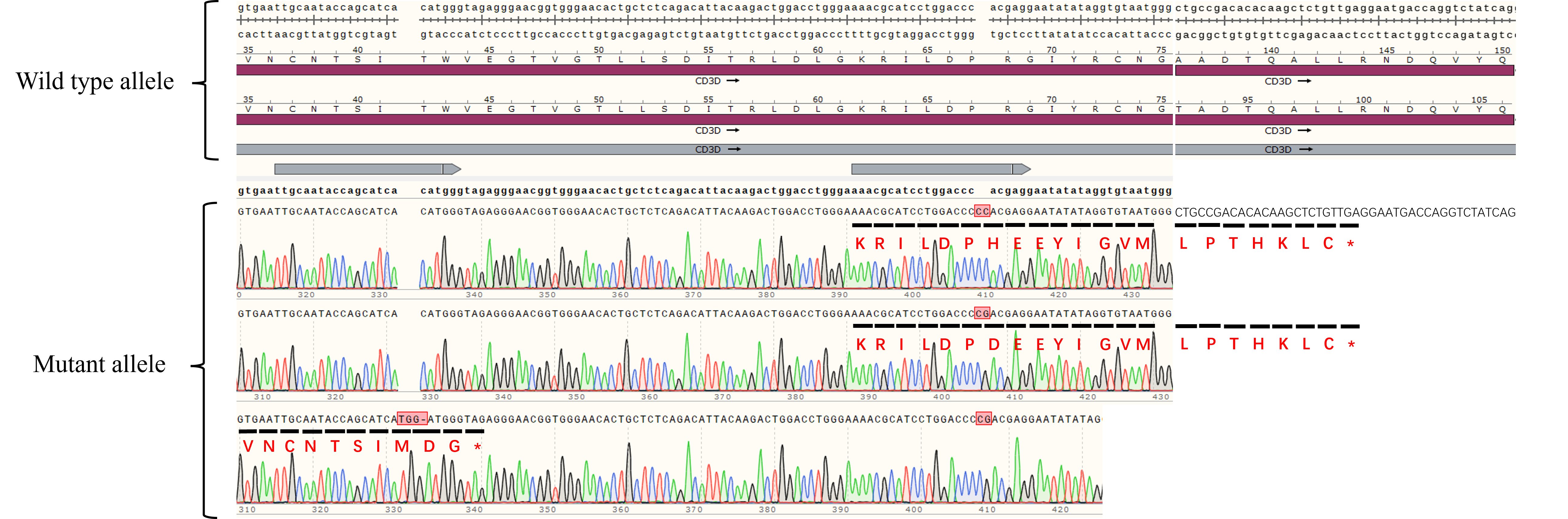
The Sanger sequencing of the Cynomolgus_CD3D CD3E Reporter Jurkat(CD3DE KO) Cell Line(Cat. GM-C45147) showed successful knockout of Human CD3D.
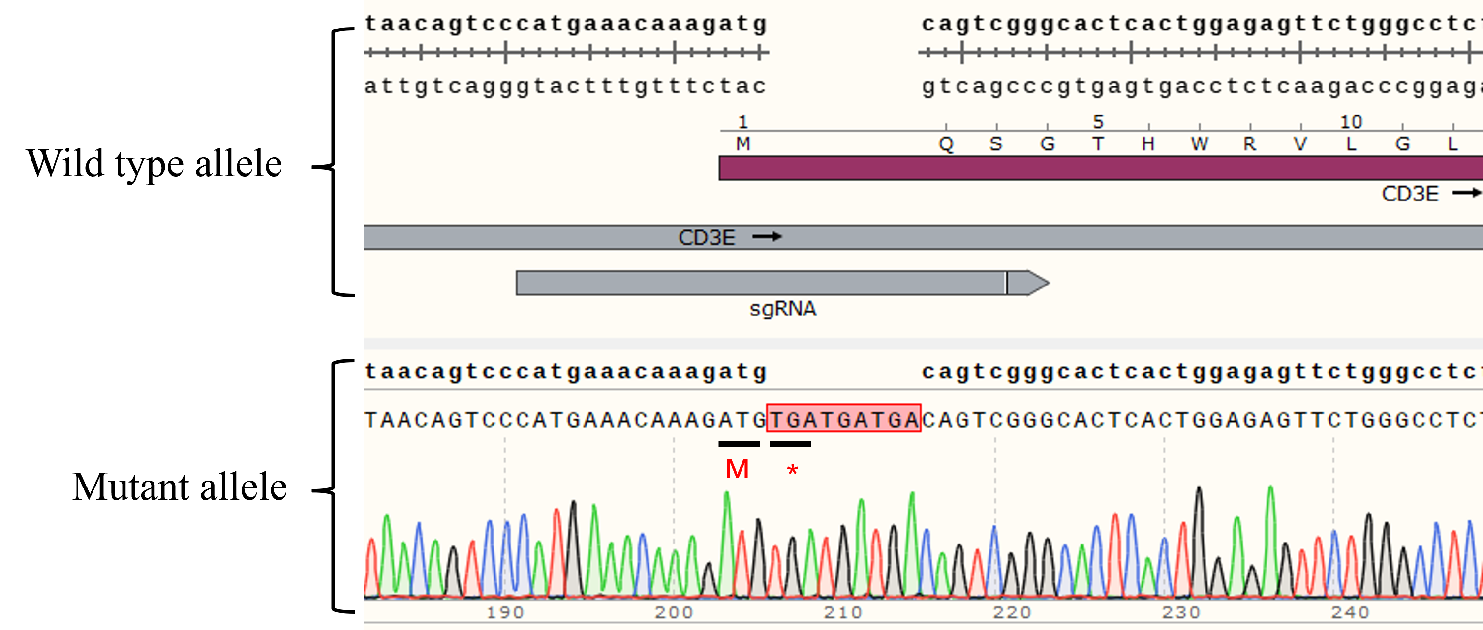
The Sanger sequencing of the Cynomolgus_CD3D CD3E Reporter Jurkat(CD3DE KO) Cell Line(Cat. GM-C45147) showed successful knockout of Human CD3E.
Specifications
Cat. No GM-C45147
Product Cynomolgus_CD3D CD3E Reporter Jurkat(CD3DE KO) Cell Line
Product Format 1 vial of frozen cells
Quantity 5E6 Cells per vial,1 mL
Storage Conditions Liquid nitrogen immediately upon receipt
Recovery Medium RPMI 1640+10% FBS+1% P.S
Growth medium RPMI 1640+10% FBS+1% P.S+3.5 μg/mL Blasticidin+400 μg/mL G418+0.75 μg/mL Puromycin
Note None
Freezing Medium 90% FBS+10% DMSO
Growth properties Suspension
Growth Conditions 37°C, 5% CO₂
Safety considerations Biosafety Level 2
Mycoplasma Testing The cell line has been screened to confirm the absence of Mycoplasma species.
Cat. No GM-C45147
Product Cynomolgus_CD3D CD3E Reporter Jurkat(CD3DE KO) Cell Line
Product Format 1 vial of frozen cells
Quantity 5E6 Cells per vial,1 mL
Storage Conditions Liquid nitrogen immediately upon receipt
Recovery Medium RPMI 1640+10% FBS+1% P.S
Growth medium RPMI 1640+10% FBS+1% P.S+3.5 μg/mL Blasticidin+400 μg/mL G418+0.75 μg/mL Puromycin
Note None
Freezing Medium 90% FBS+10% DMSO
Growth properties Suspension
Growth Conditions 37°C, 5% CO₂
Safety considerations Biosafety Level 2
Mycoplasma Testing The cell line has been screened to confirm the absence of Mycoplasma species.
Materials
Reagent Ordering Information
RPMI 1640 gibco/C11875500BT
Fetal Bovine Serum ExCell/FSP500
Pen/Strep Thermo/15140-122
Blasticidin Genomeditech/GM-040404
G418 Genomeditech/GM-040402
Puromycin Genomeditech/GM-040401
PMA/TPA Beyotime/S1819
Ionomycin MCE/HY-13434
Anti-CD3 hIgG1 Antibody(CH2527) Genomeditech/GM-33037AB
Anti-CD3 mIgG1 Antibody(SP34-2) Genomeditech/GM-87919AB
Anti-CD3 epsilon hIgG1 Antibody  Genomeditech/GM-51478AB
GMOne-Step 2.0 Luciferase Reporter Gene Assay Kit Genomeditech/GM-040513
Reagent Ordering Information
RPMI 1640 gibco/C11875500BT
Fetal Bovine Serum ExCell/FSP500
Pen/Strep Thermo/15140-122
Blasticidin Genomeditech/GM-040404
G418 Genomeditech/GM-040402
Puromycin Genomeditech/GM-040401
PMA/TPA Beyotime/S1819
Ionomycin MCE/HY-13434
Anti-CD3 hIgG1 Antibody(CH2527) Genomeditech/GM-33037AB
Anti-CD3 mIgG1 Antibody(SP34-2) Genomeditech/GM-87919AB
Anti-CD3 epsilon hIgG1 Antibody  Genomeditech/GM-51478AB
GMOne-Step 2.0 Luciferase Reporter Gene Assay Kit Genomeditech/GM-040513
Cell Culture

Cell Recovery

Recovery Medium: RPMI 1640+10% FBS+1% P.S

To insure the highest level of viability, thaw the vial and initiate the culture as soon as possible upon receipt. If upon arrival, continued storage of the frozen culture is necessary, it should be stored in liquid nitrogen vapor phase and not at -70°C. Storage at -70°C will result in loss of viability.

a)       Thaw the vial by gentle agitation in a 37°C water bath. To reduce the possibility of contamination, keep the O-ring and cap out of the water. Thawing should be rapid (approximately 2 - 3 minutes).

b)       Remove the vial from the water bath as soon as the contents are thawed, and decontaminate by dipping in or spraying with 70% ethanol. All of the operations from this point on should be carried out under strict aseptic conditions.

c)       Transfer the vial contents to a centrifuge tube containing 5.0 mL complete culture medium. And spin at approximately 176 x g for 5 minutes. Discard supernatant.

d)       Resuspend cell pellet with the recommended complete medium. And dispense the suspension into 1 - 2 T-25 culture flasks.

e)       Incubate the culture at 37°C in a suitable incubator. A 5% CO₂ in air atmosphere is recommended if using the medium described on this product sheet.

Cell Freezing

Freezing Medium: 90% FBS+10% DMSO

a)          Centrifuge at 176 x g for 3 minutes to collect cells.

b)         Resuspend the cells in pre-cooled freezing medium and adjust the cell density to 5E6 cells/mL.

c)          Aliquot 1 mL into each vial.

d)         Place the vial in a controlled-rate freezing container and store at -80°C for at least 1 day, then transfer to liquid nitrogen as soon as possible.

Cell passage

Growth medium: RPMI 1640+10% FBS+1% P.S+3.5 μg/mL Blasticidin+400 μg/mL G418+0.75 μg/mL Puromycin

Approximately 48-72 hours after the initial thawing, the cells can be passaged for the first time. After this initial passage, the culture medium can be adjusted to growth medium supplemented with antibiotics. If cells are not passaged within 48 hours, it is recommended to add some fresh recovery medium and place the flask horizontally.

a)          When the cell density reaches 1.5 - 2E6 cells/mL, subculture the cells. Do not allow the cell density to exceed 2E6 cells/mL.

b)         It is recommended to use T-25 flasks for subculturing.

c)          These cells are suspension cells, and it is recommended to use the "half-medium change" method to maintain optimal cell conditions during passaging.

d)         During passaging, you can directly add fresh growth medium to the culture flask, gently pipette to resuspend the cells, and then transfer the cell suspension to a new T-25 flask for continued culture.

Subcultivation Ratio: Maintain cultures at a cell concentraion between 3E5 and 1E6 viable cells/mL.

Medium Renewal: Every 2 to 3 days

Notes

a)          These cells are sensitive to density, so please ensure that the cell density is maintained within an appropriate range during culture and subculturing.

b)         During the first passage, pay attention to the nutrient supply; if not subculturing, make sure to add fresh recovery medium every other day as needed.

Cell Recovery

Recovery Medium: RPMI 1640+10% FBS+1% P.S

To insure the highest level of viability, thaw the vial and initiate the culture as soon as possible upon receipt. If upon arrival, continued storage of the frozen culture is necessary, it should be stored in liquid nitrogen vapor phase and not at -70°C. Storage at -70°C will result in loss of viability.

a)       Thaw the vial by gentle agitation in a 37°C water bath. To reduce the possibility of contamination, keep the O-ring and cap out of the water. Thawing should be rapid (approximately 2 - 3 minutes).

b)       Remove the vial from the water bath as soon as the contents are thawed, and decontaminate by dipping in or spraying with 70% ethanol. All of the operations from this point on should be carried out under strict aseptic conditions.

c)       Transfer the vial contents to a centrifuge tube containing 5.0 mL complete culture medium. And spin at approximately 176 x g for 5 minutes. Discard supernatant.

d)       Resuspend cell pellet with the recommended complete medium. And dispense the suspension into 1 - 2 T-25 culture flasks.

e)       Incubate the culture at 37°C in a suitable incubator. A 5% CO₂ in air atmosphere is recommended if using the medium described on this product sheet.

Cell Freezing

Freezing Medium: 90% FBS+10% DMSO

a)          Centrifuge at 176 x g for 3 minutes to collect cells.

b)         Resuspend the cells in pre-cooled freezing medium and adjust the cell density to 5E6 cells/mL.

c)          Aliquot 1 mL into each vial.

d)         Place the vial in a controlled-rate freezing container and store at -80°C for at least 1 day, then transfer to liquid nitrogen as soon as possible.

Cell passage

Growth medium: RPMI 1640+10% FBS+1% P.S+3.5 μg/mL Blasticidin+400 μg/mL G418+0.75 μg/mL Puromycin

Approximately 48-72 hours after the initial thawing, the cells can be passaged for the first time. After this initial passage, the culture medium can be adjusted to growth medium supplemented with antibiotics. If cells are not passaged within 48 hours, it is recommended to add some fresh recovery medium and place the flask horizontally.

a)          When the cell density reaches 1.5 - 2E6 cells/mL, subculture the cells. Do not allow the cell density to exceed 2E6 cells/mL.

b)         It is recommended to use T-25 flasks for subculturing.

c)          These cells are suspension cells, and it is recommended to use the "half-medium change" method to maintain optimal cell conditions during passaging.

d)         During passaging, you can directly add fresh growth medium to the culture flask, gently pipette to resuspend the cells, and then transfer the cell suspension to a new T-25 flask for continued culture.

Subcultivation Ratio: Maintain cultures at a cell concentraion between 3E5 and 1E6 viable cells/mL.

Medium Renewal: Every 2 to 3 days

Notes

a)          These cells are sensitive to density, so please ensure that the cell density is maintained within an appropriate range during culture and subculturing.

b)         During the first passage, pay attention to the nutrient supply; if not subculturing, make sure to add fresh recovery medium every other day as needed.

Related products
CD28
H_CD28 Reporter Jurkat Cell Line Cynomolgus_CD28 CHO-K1 Cell Line H_CD28 CHO-K1 Cell Line
H_CD28 HEK-293 Cell Line H_CD28 KO Jurkat Cell Line
Anti-CD28 hIgG4 Antibody(FR104) Anti-H_CD28 hIgG4 Antibody Anti-mouse CD28 Syrian Hamster IgG2 Antibody(37. 51)
Human CD28 Protein; His Tag    
CD19
H_CD19 KO Raji Cell Line Cynomolgus_CD19 CHO-K1 Cell Line Cynomolgus_CD19 HEK-293 Cell Line
H_CD19 CHO-K1 Cell line H_CD19 HEK-293 Cell Line Mouse_CD19 CHO-K1 Cell Line
Anti-CD19 hIgG1 Reference Antibody (Loncbio) Anti-CD19×CD3 hIgG1 Antibody[PIT-565(CD58 K34A)] Anti-H_CD19 hIgG1/hIgG2 Antibody
CD3
Jurkat CD3-BsAb Reporter Cell Line Cynomolgus_CD3 HEK-293 Cell Line Cynomolgus_CD3E(Membrane Bound ECD) CHO-K1 Cell Line
H_CD3 CHO-K1 Cell Line H_CD3 HEK-293 Cell Line H_CD3(TCR V2) CHO-K1 Cell Line
H_CD3(TCR V2) HEK-293 Cell Line H_CD3(TCRαβ delta V) HEK-293 Cell Line H_CD3D CD3E KO Jurkat Cell Line
H_CD3E KO Jurkat Cell Line H_CD3E(Membrane Bound ECD) CHO-K1 Cell Line Mouse_CD3 HEK-293 Cell Line
Anti-CD19×CD3 hIgG1 Antibody[PIT-565(CD58 K34A)] Anti-CD3 epsilon hIgG1 Antibody  Anti-CD3 hIgG1 Antibody(CH2527)
Anti-CD3×CD20 hIgG1 Bispecific Antibody (Epcobio) Anti-CD3×FCRL5 hIgG1 Bispecific Antibody Anti-CD3E×BCMA hIgG4 Reference Antibody (Tecbio)
Anti-CD3E×DLL3 hIgG1 Bispecific Antibody(Tarlabio) Anti-CD3E×MUC17 hIgG1 Bispecific Antibody(Vepsitbio) Anti-mouse CD3ε mIgG2a Antibody(145-2C11)
CD2
H_CD2 KO Jurkat(CD3-) Cell Line CD3-CD2-tsAb Reporter Jurkat(CD58 KO) Cell Line Cynomolgus_CD2 CHO-K1 Cell Line
H_CD2 CHO-K1 Cell Line
Anti-CD19×CD3 hIgG1 Antibody[PIT-565(CD58 K34A)] Anti-CD2 hIgG1 Antibody(BTI-322)
Human CD2 Protein; hFc Tag Human CD2 Protein; His Tag  
For laboratory research use only. Direct human use, including taking orally and injection and clinical use are forbidden.
Cynomolgus_CD3D CD3E Reporter Jurkat(CD3DE KO) Cell
Cat. No.
GM-C45147
Size
2 vial
Quote
Description
Data Display
Specifications
Materials
Cell Culture
Related products
Description
CD3D and CD3E form the CD3δε heterodimer, a core subunit of the TCR‑CD3 complex essential for proper assembly and surface expression. Upon TCR engagement, their cytoplasmic ITAMs are phosphorylated, triggering downstream signaling that drives T‑cell activation, proliferation, and effector functions. CD3D is critical for thymocyte development, while CD3E regulates receptor internalization. As a major target in bispecific antibody development (over 400 active programs), most CD3‑targeting antibodies recognize CD3ε and mediate antitumor effects through T‑cell engagement. Therefore, a cell model expressing cynomolgus CD3 is valuable for preclinical cross‑reactivity and pharmacodynamic assessment.

Cynomolgus_CD3D CD3E Reporter Jurkat(CD3DE KO) Cell Line is a clonal stable cell line that knockout CD3D/E and constitutive expression of Cynomolgus_CD3D/E gene, along with signal-dependent expression of a luciferase reporter gene. When drugs capable of binding and activating cynomolgus CD3 are added, the TCR‑CD3 complex becomes cross‑linked, triggering downstream signaling pathways that drive luciferase expression. The luciferase readout reflects the extent of pathway activation and can be used to evaluate the agonistic activity or functional binding of candidate drugs to cynomolgus CD3.

CD3D and CD3E form the CD3δε heterodimer, a core subunit of the TCR‑CD3 complex essential for proper assembly and surface expression. Upon TCR engagement, their cytoplasmic ITAMs are phosphorylated, triggering downstream signaling that drives T‑cell activation, proliferation, and effector functions. CD3D is critical for thymocyte development, while CD3E regulates receptor internalization. As a major target in bispecific antibody development (over 400 active programs), most CD3‑targeting antibodies recognize CD3ε and mediate antitumor effects through T‑cell engagement. Therefore, a cell model expressing cynomolgus CD3 is valuable for preclinical cross‑reactivity and pharmacodynamic assessment.

Cynomolgus_CD3D CD3E Reporter Jurkat(CD3DE KO) Cell Line is a clonal stable cell line that knockout CD3D/E and constitutive expression of Cynomolgus_CD3D/E gene, along with signal-dependent expression of a luciferase reporter gene. When drugs capable of binding and activating cynomolgus CD3 are added, the TCR‑CD3 complex becomes cross‑linked, triggering downstream signaling pathways that drive luciferase expression. The luciferase readout reflects the extent of pathway activation and can be used to evaluate the agonistic activity or functional binding of candidate drugs to cynomolgus CD3.

Data Display
Signaling Pathway
Response to Anti-CD3 hIgG1 Antibody(CH2527). The Cynomolgus_CD3D CD3E Reporter Jurkat(CD3DE KO) Cell Line (Cat. GM-C45147) at a concentration of 5E4 cells/well (96-well format) was stimulated with serial dilutions of Anti-CD3 hIgG1 Antibody(CH2527) (Cat. GM-33037AB) in assay buffer (RPMI 1640+1% FBS+1% P.S) for 6 hours. The firefly luciferase activity was measured using the Luciferase Reporter Assay Kit (Genomeditech). The maximum induction fold was approximately [126.1]. Data are shown by drug mass concentration.
Response to Anti-CD3 mIgG1 Antibody(SP34-2). The Cynomolgus_CD3D CD3E Reporter Jurkat(CD3DE KO) Cell Line (Cat. GM-C45147) at a concentration of 5E4 cells/well (96-well format) was stimulated with serial dilutions of Anti-CD3 mIgG1 Antibody(SP34-2) (Cat. GM-87919AB) and Anti-CD3 epsilon Antibody (Cat. GM-51478AB) in assay buffer (RPMI 1640+1% FBS+1% P.S) for 6 hours. The firefly luciferase activity was measured using the Luciferase Reporter Assay Kit (Genomeditech). The firefly luciferase activity was measured using the Luciferase Reporter Assay Kit (Genomeditech). The maximum induction fold was approximately [136.6]. Data are shown by drug mass concentration.
Response to PMA/TPA. The Cynomolgus_CD3D CD3E Reporter Jurkat(CD3DE KO) Cell Line (Cat. GM-C45147) at a concentration of 5E4 cells/well (96-well format) was stimulated with serial dilutions of PMA/TPA (Beyotime/S1819) and 500nM Ionomycin (MCE/HY-13434) in assay buffer (RPMI 1640+1% FBS+1% P.S) for 6 hours. The firefly luciferase activity was measured using the Luciferase Reporter Assay Kit (Genomeditech). The maximum induction fold was approximately [9.7]. Data are shown by drug molar concentration.
Expression
Cynomolgus_CD3D CD3E Reporter Jurkat(CD3DE KO) Cell Line (Cat. GM-C45147) was determined by flow cytometry using Anti-CD3 hIgG1 Antibody(CH2527)(Cat. GM-33037AB).
Genotype Verification
Cynomolgus_CD3D CD3E Reporter Jurkat(CD3DE KO) Cell Line (Cat. GM-C45147) was determined by flow cytometry using Anti-CD3 epsilon hIgG1 Antibody (Cat. GM-51478AB).(The antibody does not recognize Cynomolgus.)
The Sanger sequencing of the Cynomolgus_CD3D CD3E Reporter Jurkat(CD3DE KO) Cell Line(Cat. GM-C45147) showed successful knockout of Human CD3D.
The Sanger sequencing of the Cynomolgus_CD3D CD3E Reporter Jurkat(CD3DE KO) Cell Line(Cat. GM-C45147) showed successful knockout of Human CD3E.
Specifications
Cat. No GM-C45147
Product Cynomolgus_CD3D CD3E Reporter Jurkat(CD3DE KO) Cell Line
Product Format 1 vial of frozen cells
Quantity 5E6 Cells per vial,1 mL
Storage Conditions Liquid nitrogen immediately upon receipt
Recovery Medium RPMI 1640+10% FBS+1% P.S
Growth medium RPMI 1640+10% FBS+1% P.S+3.5 μg/mL Blasticidin+400 μg/mL G418+0.75 μg/mL Puromycin
Note None
Freezing Medium 90% FBS+10% DMSO
Growth properties Suspension
Growth Conditions 37°C, 5% CO₂
Safety considerations Biosafety Level 2
Mycoplasma Testing The cell line has been screened to confirm the absence of Mycoplasma species.
Cat. No GM-C45147
Product Cynomolgus_CD3D CD3E Reporter Jurkat(CD3DE KO) Cell Line
Product Format 1 vial of frozen cells
Quantity 5E6 Cells per vial,1 mL
Storage Conditions Liquid nitrogen immediately upon receipt
Recovery Medium RPMI 1640+10% FBS+1% P.S
Growth medium RPMI 1640+10% FBS+1% P.S+3.5 μg/mL Blasticidin+400 μg/mL G418+0.75 μg/mL Puromycin
Note None
Freezing Medium 90% FBS+10% DMSO
Growth properties Suspension
Growth Conditions 37°C, 5% CO₂
Safety considerations Biosafety Level 2
Mycoplasma Testing The cell line has been screened to confirm the absence of Mycoplasma species.
Materials
Reagent Ordering Information
RPMI 1640 gibco/C11875500BT
Fetal Bovine Serum ExCell/FSP500
Pen/Strep Thermo/15140-122
Blasticidin Genomeditech/GM-040404
G418 Genomeditech/GM-040402
Puromycin Genomeditech/GM-040401
PMA/TPA Beyotime/S1819
Ionomycin MCE/HY-13434
Anti-CD3 hIgG1 Antibody(CH2527) Genomeditech/GM-33037AB
Anti-CD3 mIgG1 Antibody(SP34-2) Genomeditech/GM-87919AB
Anti-CD3 epsilon hIgG1 Antibody  Genomeditech/GM-51478AB
GMOne-Step 2.0 Luciferase Reporter Gene Assay Kit Genomeditech/GM-040513
Reagent Ordering Information
RPMI 1640 gibco/C11875500BT
Fetal Bovine Serum ExCell/FSP500
Pen/Strep Thermo/15140-122
Blasticidin Genomeditech/GM-040404
G418 Genomeditech/GM-040402
Puromycin Genomeditech/GM-040401
PMA/TPA Beyotime/S1819
Ionomycin MCE/HY-13434
Anti-CD3 hIgG1 Antibody(CH2527) Genomeditech/GM-33037AB
Anti-CD3 mIgG1 Antibody(SP34-2) Genomeditech/GM-87919AB
Anti-CD3 epsilon hIgG1 Antibody  Genomeditech/GM-51478AB
GMOne-Step 2.0 Luciferase Reporter Gene Assay Kit Genomeditech/GM-040513
Cell Culture

Cell Recovery

Recovery Medium: RPMI 1640+10% FBS+1% P.S

To insure the highest level of viability, thaw the vial and initiate the culture as soon as possible upon receipt. If upon arrival, continued storage of the frozen culture is necessary, it should be stored in liquid nitrogen vapor phase and not at -70°C. Storage at -70°C will result in loss of viability.

a)       Thaw the vial by gentle agitation in a 37°C water bath. To reduce the possibility of contamination, keep the O-ring and cap out of the water. Thawing should be rapid (approximately 2 - 3 minutes).

b)       Remove the vial from the water bath as soon as the contents are thawed, and decontaminate by dipping in or spraying with 70% ethanol. All of the operations from this point on should be carried out under strict aseptic conditions.

c)       Transfer the vial contents to a centrifuge tube containing 5.0 mL complete culture medium. And spin at approximately 176 x g for 5 minutes. Discard supernatant.

d)       Resuspend cell pellet with the recommended complete medium. And dispense the suspension into 1 - 2 T-25 culture flasks.

e)       Incubate the culture at 37°C in a suitable incubator. A 5% CO₂ in air atmosphere is recommended if using the medium described on this product sheet.

Cell Freezing

Freezing Medium: 90% FBS+10% DMSO

a)          Centrifuge at 176 x g for 3 minutes to collect cells.

b)         Resuspend the cells in pre-cooled freezing medium and adjust the cell density to 5E6 cells/mL.

c)          Aliquot 1 mL into each vial.

d)         Place the vial in a controlled-rate freezing container and store at -80°C for at least 1 day, then transfer to liquid nitrogen as soon as possible.

Cell passage

Growth medium: RPMI 1640+10% FBS+1% P.S+3.5 μg/mL Blasticidin+400 μg/mL G418+0.75 μg/mL Puromycin

Approximately 48-72 hours after the initial thawing, the cells can be passaged for the first time. After this initial passage, the culture medium can be adjusted to growth medium supplemented with antibiotics. If cells are not passaged within 48 hours, it is recommended to add some fresh recovery medium and place the flask horizontally.

a)          When the cell density reaches 1.5 - 2E6 cells/mL, subculture the cells. Do not allow the cell density to exceed 2E6 cells/mL.

b)         It is recommended to use T-25 flasks for subculturing.

c)          These cells are suspension cells, and it is recommended to use the "half-medium change" method to maintain optimal cell conditions during passaging.

d)         During passaging, you can directly add fresh growth medium to the culture flask, gently pipette to resuspend the cells, and then transfer the cell suspension to a new T-25 flask for continued culture.

Subcultivation Ratio: Maintain cultures at a cell concentraion between 3E5 and 1E6 viable cells/mL.

Medium Renewal: Every 2 to 3 days

Notes

a)          These cells are sensitive to density, so please ensure that the cell density is maintained within an appropriate range during culture and subculturing.

b)         During the first passage, pay attention to the nutrient supply; if not subculturing, make sure to add fresh recovery medium every other day as needed.

Cell Recovery

Recovery Medium: RPMI 1640+10% FBS+1% P.S

To insure the highest level of viability, thaw the vial and initiate the culture as soon as possible upon receipt. If upon arrival, continued storage of the frozen culture is necessary, it should be stored in liquid nitrogen vapor phase and not at -70°C. Storage at -70°C will result in loss of viability.

a)       Thaw the vial by gentle agitation in a 37°C water bath. To reduce the possibility of contamination, keep the O-ring and cap out of the water. Thawing should be rapid (approximately 2 - 3 minutes).

b)       Remove the vial from the water bath as soon as the contents are thawed, and decontaminate by dipping in or spraying with 70% ethanol. All of the operations from this point on should be carried out under strict aseptic conditions.

c)       Transfer the vial contents to a centrifuge tube containing 5.0 mL complete culture medium. And spin at approximately 176 x g for 5 minutes. Discard supernatant.

d)       Resuspend cell pellet with the recommended complete medium. And dispense the suspension into 1 - 2 T-25 culture flasks.

e)       Incubate the culture at 37°C in a suitable incubator. A 5% CO₂ in air atmosphere is recommended if using the medium described on this product sheet.

Cell Freezing

Freezing Medium: 90% FBS+10% DMSO

a)          Centrifuge at 176 x g for 3 minutes to collect cells.

b)         Resuspend the cells in pre-cooled freezing medium and adjust the cell density to 5E6 cells/mL.

c)          Aliquot 1 mL into each vial.

d)         Place the vial in a controlled-rate freezing container and store at -80°C for at least 1 day, then transfer to liquid nitrogen as soon as possible.

Cell passage

Growth medium: RPMI 1640+10% FBS+1% P.S+3.5 μg/mL Blasticidin+400 μg/mL G418+0.75 μg/mL Puromycin

Approximately 48-72 hours after the initial thawing, the cells can be passaged for the first time. After this initial passage, the culture medium can be adjusted to growth medium supplemented with antibiotics. If cells are not passaged within 48 hours, it is recommended to add some fresh recovery medium and place the flask horizontally.

a)          When the cell density reaches 1.5 - 2E6 cells/mL, subculture the cells. Do not allow the cell density to exceed 2E6 cells/mL.

b)         It is recommended to use T-25 flasks for subculturing.

c)          These cells are suspension cells, and it is recommended to use the "half-medium change" method to maintain optimal cell conditions during passaging.

d)         During passaging, you can directly add fresh growth medium to the culture flask, gently pipette to resuspend the cells, and then transfer the cell suspension to a new T-25 flask for continued culture.

Subcultivation Ratio: Maintain cultures at a cell concentraion between 3E5 and 1E6 viable cells/mL.

Medium Renewal: Every 2 to 3 days

Notes

a)          These cells are sensitive to density, so please ensure that the cell density is maintained within an appropriate range during culture and subculturing.

b)         During the first passage, pay attention to the nutrient supply; if not subculturing, make sure to add fresh recovery medium every other day as needed.

Related products
CD28
H_CD28 Reporter Jurkat Cell Line Cynomolgus_CD28 CHO-K1 Cell Line H_CD28 CHO-K1 Cell Line
H_CD28 HEK-293 Cell Line H_CD28 KO Jurkat Cell Line
Anti-CD28 hIgG4 Antibody(FR104) Anti-H_CD28 hIgG4 Antibody Anti-mouse CD28 Syrian Hamster IgG2 Antibody(37. 51)

Human CD28 Protein; His Tag    
CD19
H_CD19 KO Raji Cell Line Cynomolgus_CD19 CHO-K1 Cell Line Cynomolgus_CD19 HEK-293 Cell Line
H_CD19 CHO-K1 Cell line H_CD19 HEK-293 Cell Line Mouse_CD19 CHO-K1 Cell Line
Anti-CD19 hIgG1 Reference Antibody (Loncbio) Anti-CD19×CD3 hIgG1 Antibody[PIT-565(CD58 K34A)] Anti-H_CD19 hIgG1/hIgG2 Antibody
CD3
Jurkat CD3-BsAb Reporter Cell Line Cynomolgus_CD3 HEK-293 Cell Line Cynomolgus_CD3E(Membrane Bound ECD) CHO-K1 Cell Line
H_CD3 CHO-K1 Cell Line H_CD3 HEK-293 Cell Line H_CD3(TCR V2) CHO-K1 Cell Line
H_CD3(TCR V2) HEK-293 Cell Line H_CD3(TCRαβ delta V) HEK-293 Cell Line H_CD3D CD3E KO Jurkat Cell Line
H_CD3E KO Jurkat Cell Line H_CD3E(Membrane Bound ECD) CHO-K1 Cell Line Mouse_CD3 HEK-293 Cell Line
Anti-CD19×CD3 hIgG1 Antibody[PIT-565(CD58 K34A)] Anti-CD3 epsilon hIgG1 Antibody  Anti-CD3 hIgG1 Antibody(CH2527)
Anti-CD3×CD20 hIgG1 Bispecific Antibody (Epcobio) Anti-CD3×FCRL5 hIgG1 Bispecific Antibody Anti-CD3E×BCMA hIgG4 Reference Antibody (Tecbio)
Anti-CD3E×DLL3 hIgG1 Bispecific Antibody(Tarlabio) Anti-CD3E×MUC17 hIgG1 Bispecific Antibody(Vepsitbio) Anti-mouse CD3ε mIgG2a Antibody(145-2C11)
CD2
H_CD2 KO Jurkat(CD3-) Cell Line CD3-CD2-tsAb Reporter Jurkat(CD58 KO) Cell Line Cynomolgus_CD2 CHO-K1 Cell Line
H_CD2 CHO-K1 Cell Line
Anti-CD19×CD3 hIgG1 Antibody[PIT-565(CD58 K34A)] Anti-CD2 hIgG1 Antibody(BTI-322)
Human CD2 Protein; hFc Tag Human CD2 Protein; His Tag  
Contact
Us
Message Inquiry
reset
submit
Message
Message Inquiry
reset
submit